


Introduction: The Vital Importance of Knowing the Heart’s Structure
Understanding the heart is far more than an academic pursuit; it is a critical foundation for grasping how life itself is sustained. The human heart, a muscular organ no larger than a clenched fist, plays an indispensable role in maintaining the flow of blood—and thus oxygen and nutrients—throughout the body. While heart disease remains the leading cause of death globally, increased awareness of cardiac structure and function can empower individuals to take charge of their health. This article explores the heart through a visual and anatomical lens, offering labeled images of the heart that reveal how its intricate parts work together to sustain life. In doing so, it promotes a deeper, science-backed understanding of cardiovascular health and its real-world implications.
You may also like: 5 Ways to Keep Your Heart Healthy and Prevent Cardiovascular Disease
This article is designed to serve both medically curious readers and health professionals alike. With a college graduate-level audience in mind, the information provided here is thoroughly researched, scientifically accurate, and free of sensationalism. Using labeled images of the heart as a guide, this article will not only describe the heart’s anatomy in rich detail but will also illuminate how this structure underpins function. Each section builds on the previous, gradually expanding the reader’s comprehension through both text and visual aids.
The Heart as a Pump: Basic Structure and Circulatory Function
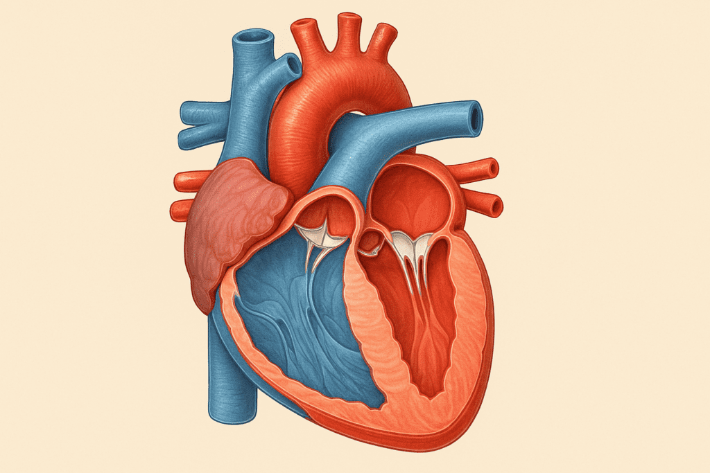
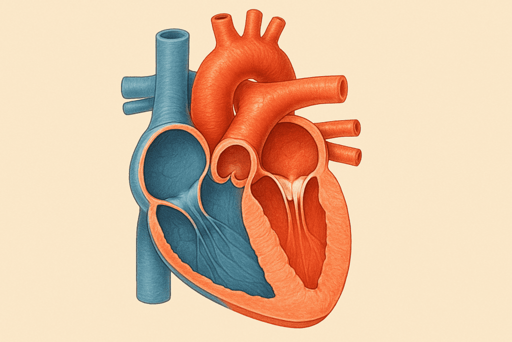
The human heart serves as a dual pump system, divided into left and right sides that each perform distinct yet interconnected functions. The right side receives deoxygenated blood from the body and pumps it into the lungs, where carbon dioxide is exchanged for oxygen. Conversely, the left side of the heart receives oxygen-rich blood from the lungs and propels it into systemic circulation to nourish organs and tissues. Each side contains two chambers: an atrium and a ventricle. The atria receive blood, while the ventricles are responsible for pumping it out. Understanding this division is critical for grasping why certain cardiac diseases impact one side of the heart more than the other.
Labeled images of the heart clearly illustrate this four-chambered structure. They help clarify how blood moves sequentially through the superior and inferior vena cava into the right atrium, through the tricuspid valve into the right ventricle, and on to the pulmonary artery and lungs. After oxygenation, blood travels through the pulmonary veins to the left atrium, down through the mitral valve into the left ventricle, and finally out through the aorta. Without a clear visualization, such as a labeled diagram, this complex flow can easily become abstract. Therefore, understanding the heart through labeled images of the heart that reveal its structure and function is an essential step in cardiac education.
Major Blood Vessels: Gateways of Circulation
No discussion of cardiac anatomy is complete without examining the major blood vessels that interface directly with the heart. These vessels include the aorta, pulmonary arteries and veins, and the venae cavae. Each plays a pivotal role in either oxygenating blood or delivering it throughout the body. The aorta, for instance, is the largest artery in the body and serves as the main conduit for oxygen-rich blood leaving the left ventricle. The superior and inferior vena cava carry deoxygenated blood back to the right atrium from the upper and lower body, respectively.
When viewed in labeled images of the heart that reveal its structure and function, these vessels appear as major highways into and out of the organ. The relationship between vessel size, location, and function becomes much easier to comprehend through visual representation. These diagrams also highlight how arterial and venous systems are distinct yet interdependent. For example, while the pulmonary artery carries deoxygenated blood away from the heart, the pulmonary veins uniquely carry oxygenated blood back into it. This is a reversal of the standard pattern seen elsewhere in the body, and understanding it can prevent misconceptions that are common even among students in health sciences.
Valves of the Heart: Guardians of Unidirectional Flow
The heart’s valves ensure that blood flows in only one direction, preventing any backward leakage. There are four main valves: the tricuspid, pulmonary, mitral, and aortic valves. Each valve opens and closes in synchrony with the heartbeat, driven by pressure differences across its leaflets. Malfunction of any of these valves—such as in valve stenosis or regurgitation—can lead to serious health consequences, including heart failure and stroke.
Labeled heart diagrams make it easier to understand where these valves are located and how they interact with surrounding chambers. By examining labeled images of the heart that reveal its structure and function, we can visualize the positioning and role of each valve. For instance, the mitral valve is situated between the left atrium and left ventricle, and its failure to close completely can lead to backflow and volume overload. Understanding valve anatomy not only clarifies clinical terminology—like “mitral valve prolapse”—but also underscores the delicate coordination required for efficient cardiac output.
Myocardium, Endocardium, and Pericardium: The Heart’s Three-Layered Wall
The heart wall consists of three distinct layers, each contributing to overall function. The innermost layer, the endocardium, lines the heart’s chambers and valves, offering a smooth surface for blood flow and protecting against thrombosis. The myocardium, the thick muscular middle layer, is responsible for the contractile force that propels blood throughout the body. Lastly, the pericardium—a double-walled sac surrounding the heart—provides structural support and minimizes friction between the heart and nearby organs.
When labeled images of the heart are used to reveal its structure and function, the layers of the heart wall can be seen in cross-sectional views. These images are indispensable for medical students learning about myocardial infarction, where damage is typically localized to the myocardium. Similarly, conditions like pericarditis become more intelligible when one can visualize the inflamed pericardial sac. The ability to directly see these layers helps bridge the gap between textbook definitions and clinical realities.
The Cardiac Conduction System: Wiring the Heart’s Electrical Signals
The heart does not contract at random—it follows a highly coordinated electrical rhythm governed by specialized tissues. The sinoatrial (SA) node, often called the heart’s natural pacemaker, initiates each heartbeat by generating an electrical impulse. This impulse travels through the atria, causing them to contract, before reaching the atrioventricular (AV) node. The AV node then relays the signal down the bundle of His and into the Purkinje fibers, leading to synchronized contraction of the ventricles.
Visual aids are particularly helpful in illustrating this invisible yet vital system. Labeled images of the heart that reveal its structure and function often include pathways of the conduction system, clarifying how impulses travel and where disruptions can occur. Such images demystify arrhythmias, heart blocks, and the role of pacemakers. They show, for example, why atrial fibrillation originates above the AV node and how this affects ventricular response. With the rise of electrophysiology as a cardiology subspecialty, such understanding is more relevant than ever.
Coronary Circulation: Feeding the Heart Itself
While the heart pumps blood to the rest of the body, it also requires its own dedicated blood supply. This is provided by the coronary arteries, which branch off from the base of the aorta. The right and left coronary arteries and their branches deliver oxygenated blood to the myocardium. When these arteries become blocked—most commonly due to atherosclerosis—a myocardial infarction or “heart attack” can occur. The resulting damage can severely impair the heart’s ability to function.
Labeled diagrams are critical tools in understanding how coronary arteries wrap around the heart and which regions they supply. When we explore labeled images of the heart that reveal its structure and function, the course of the right coronary artery versus the left anterior descending artery becomes much easier to distinguish. This has practical importance in understanding EKG patterns and interpreting cardiac catheterization results. For example, ST-segment elevation in specific EKG leads often correlates with occlusion in distinct coronary branches, a relationship best understood when paired with anatomical visuals.
Cardiac Chambers: Functional and Structural Insights
Each chamber of the heart serves a unique and vital role. The right atrium receives deoxygenated blood from systemic veins, while the right ventricle pumps it into the pulmonary circulation. The left atrium receives oxygenated blood from the lungs, and the left ventricle propels it into systemic arteries. The left ventricle is particularly muscular, as it must generate sufficient pressure to deliver blood throughout the entire body. This difference in muscle thickness is often apparent in labeled heart cross-sections.
Understanding the heart through labeled images of the heart that reveal its structure and function allows for direct comparison between chambers. For instance, a diagram may show the hypertrophy of the left ventricle in a patient with chronic hypertension or the dilation of the right ventricle in pulmonary hypertension. Visualizing chamber size and wall thickness helps in interpreting echocardiograms and other imaging modalities. Furthermore, these structural features are key to understanding heart failure classifications, such as reduced ejection fraction versus preserved ejection fraction.
Frequently Asked Questions: Understanding the Heart Through Labeled Visuals
What makes labeled heart images more effective than written descriptions alone?
While text-based explanations can be highly informative, the brain often processes visual data more efficiently. A picture of heart labeled with anatomical precision allows learners to form spatial associations, making it easier to remember the heart’s structure. This visual reinforcement is particularly helpful when studying complex concepts like blood flow pathways or valve positioning. In clinical settings, heart anatomy images also facilitate patient education, helping individuals understand their diagnosis and treatment options. The combination of clear visuals and concise annotations in images of the heart labeled provides an immersive learning experience that written descriptions alone cannot match.
How do labeled images enhance the diagnosis of heart conditions?
Medical imaging techniques such as echocardiograms, MRIs, and CT scans often produce raw anatomical data that can be overwhelming without proper context. When paired with a picture of heart labeled accurately, clinicians can compare patient-specific images to standard anatomical references. This comparative method aids in identifying abnormalities like chamber enlargement, valve dysfunction, or congenital defects. Furthermore, understanding these discrepancies in the context of heart anatomy images helps ensure more accurate diagnoses and better-informed treatment decisions. Physicians often rely on images of the heart labeled to explain procedures such as angioplasty or valve replacement in a way that resonates with patients.
Can labeled heart diagrams help patients communicate more effectively with doctors?
Yes, absolutely. Many patients feel intimidated by medical jargon, which can hinder meaningful dialogue during consultations. A picture of heart labeled with clear terms creates a visual bridge between physician and patient, enabling better two-way communication. When patients can point to a specific part on heart anatomy images, they gain confidence in expressing symptoms or concerns. This improved clarity can lead to earlier diagnoses, stronger patient engagement, and increased adherence to treatment plans. In fact, using images of the heart labeled in patient discussions has been shown to enhance health literacy, particularly among those without a strong background in science.
How are labeled heart images used in surgical planning and training?
Surgical precision depends heavily on a deep understanding of individual anatomical variation. Surgeons often consult highly detailed heart anatomy images prior to operations, sometimes even superimposing a picture of heart labeled over a patient’s scan to map out incision sites or assess risk. These images are also instrumental in the training of medical students and residents, who use them to simulate procedures and practice navigation through complex cardiac regions. For instance, a comprehensive set of images of the heart labeled can assist in mastering bypass graft placements or pacemaker lead insertions. By integrating these visuals into both preoperative and educational settings, medical teams improve outcomes and safety.
Do labeled heart visuals evolve with new research and technology?
They do—and quite rapidly. Advances in imaging technology, such as 3D modeling and augmented reality (AR), are revolutionizing how we view and interpret heart anatomy images. What was once a static picture of heart labeled in a textbook is now an interactive, rotating model accessible via mobile devices or virtual platforms. These modern tools not only depict standard anatomical structures but can also incorporate real-time patient data for personalized visualizations. As a result, newer images of the heart labeled often reflect the most up-to-date understanding of anatomical variants, pathologies, and functional dynamics. This evolution ensures that visual learning tools remain aligned with current scientific knowledge and clinical best practices.
Can teachers and students use labeled heart images in interdisciplinary education?
Certainly. While primarily used in medical and health science contexts, a picture of heart labeled can also be a valuable resource in fields such as biology, physiology, and even biomedical engineering. Educators across disciplines use heart anatomy images to teach circulatory principles, organ design, and mechanical modeling. For students, engaging with images of the heart labeled can foster a deeper appreciation of both anatomical complexity and biological elegance. Additionally, interdisciplinary learning becomes more dynamic when visual materials transcend traditional subject boundaries, allowing for collaborative inquiry across medicine, technology, and education.
How can labeled heart diagrams be personalized for individual learning needs?
Digital education platforms now allow users to customize their learning experiences with tools like interactive quizzes, layered visualizations, and 3D overlays. For example, a picture of heart labeled can be color-coded based on blood flow, pressure gradients, or associated diseases, depending on the learner’s focus. Students with visual learning preferences may benefit from rotating or animated heart anatomy images that show dynamic movement of valves and blood. These customized images of the heart labeled can accommodate different learning speeds and styles, empowering both self-directed learners and classroom-based students. Personalization ensures that complex topics become more approachable and tailored to individual goals.
Are there emotional or psychological benefits to using visual aids in cardiac care?
Yes, and they are often underestimated. A picture of heart labeled not only aids in comprehension but can also reduce anxiety by demystifying the heart’s inner workings. For patients newly diagnosed with cardiovascular conditions, heart anatomy images offer a sense of control and clarity in what can otherwise be an overwhelming experience. Seeing images of the heart labeled with familiar terms gives patients a tangible reference point for understanding procedures, recovery timelines, and prognosis. This visual grounding contributes to emotional reassurance and can even improve adherence to treatment protocols by reinforcing the importance of anatomical knowledge in recovery.
What role do labeled visuals play in remote or telehealth cardiac care?
In an increasingly digital healthcare landscape, telemedicine has become a vital tool for delivering cardiovascular care across distances. A picture of heart labeled becomes especially valuable in virtual settings, where tactile models or in-person demonstrations are unavailable. Providers can share high-resolution heart anatomy images via screen-sharing tools to walk patients through diagnoses, monitor disease progression, or explain interventions. These digital images of the heart labeled offer continuity of care by making virtual visits more interactive and informative. As telehealth continues to expand, integrating labeled visuals will be essential to maintaining high-quality, patient-centered care.
How can labeled heart images help prevent cardiovascular disease?
Preventative health hinges on education, and visual tools can make abstract risk factors more tangible. A picture of heart labeled with key anatomical landmarks can highlight where issues like plaque buildup or valve narrowing typically occur, encouraging early lifestyle interventions. Heart anatomy images used in public health campaigns or educational workshops can make discussions of diet, exercise, and screening more impactful. When people see images of the heart labeled with both normal and diseased states, they’re more likely to internalize the consequences of poor cardiovascular choices. In this way, labeled diagrams serve not only as teaching tools but as motivators for proactive health behavior change.
Conclusion: Why Labeled Images Are Essential for Understanding the Heart
Visual learning tools have long been central to mastering complex anatomical systems, and this is especially true for cardiac anatomy. Labeled images of the heart that reveal its structure and function serve not only as educational supplements but also as critical tools for patient education, clinical training, and medical research. They transform abstract descriptions into concrete visual understanding, making it easier to grasp the intricate architecture and physiological dynamics of the heart.
As cardiovascular disease continues to pose a significant global health burden, a deeper, more precise understanding of heart anatomy is urgently needed. From medical students to patients navigating new diagnoses, the ability to interpret and apply anatomical information can greatly enhance decision-making and health outcomes. By returning again and again to labeled images of the heart that reveal its structure and function, we reinforce our understanding, sharpen our clinical intuition, and build a more compassionate, evidence-based approach to cardiovascular care.
In the end, the heart is more than an organ—it is a marvel of biological engineering. And through labeled images that bring its design to life, we are granted not only the chance to understand but also to appreciate the profound intricacies that sustain our every heartbeat.
human heart anatomy, cardiovascular system diagram, internal heart structure, parts of the human heart, cardiac anatomy guide, visual anatomy of the heart, circulatory system organs, how the heart works, heart health education, cardiac imaging tools, anatomy for medical students, heart disease awareness, educational heart diagrams, function of heart chambers, anatomy of blood vessels, valve function in the heart, cardiac muscle structure, coronary circulation explained, understanding heart valves, heart education for patients
Further Reading:
Heart Anatomy, Function, and Blood Circulation
Towards new understanding of the heart structure and function
Disclaimer
The information contained in this article is provided for general informational purposes only and is not intended to serve as medical, legal, or professional advice. While MedNewsPedia strives to present accurate, up-to-date, and reliable content, no warranty or guarantee, expressed or implied, is made regarding the completeness, accuracy, or adequacy of the information provided. Readers are strongly advised to seek the guidance of a qualified healthcare provider or other relevant professionals before acting on any information contained in this article. MedNewsPedia, its authors, editors, and contributors expressly disclaim any liability for any damages, losses, or consequences arising directly or indirectly from the use, interpretation, or reliance on any information presented herein. The views and opinions expressed in this article are those of the author(s) and do not necessarily reflect the official policies or positions of MedNewsPedia.