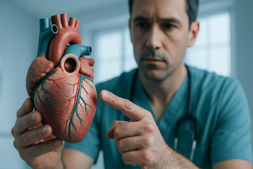
Understanding the Anatomy of the Human Heart
The human heart is one of the most intricate and vital organs in the body, responsible for circulating blood, delivering oxygen and nutrients, and maintaining the pressure that sustains life. At the top of this powerful muscular pump is a distinctive area where several major structures converge—what many refer to colloquially as the “circle thing at the top of the heart.” While this informal phrase may sound imprecise, it generally refers to the base of the heart, a critical anatomical zone where the great vessels, such as the aorta and pulmonary artery, attach. Understanding the form and function of this region is key to appreciating how the heart sustains systemic and pulmonary circulation.
You may also like: 5 Ways to Keep Your Heart Healthy and Prevent Cardiovascular Disease
The base of the heart forms its broad superior surface, contrasting with the pointed inferior apex. This area houses not only the openings of the major arteries and veins but also the upper chambers of the heart, or atria, which play an essential role in receiving blood and regulating its flow into the ventricles. The base is fixed in position due to its connection with the large vessels and is located near the second intercostal space behind the sternum. Recognizing the anatomical distinction between the base and apex of the heart helps clinicians accurately interpret imaging and physical exam findings.
What Is the Circle Thing at the Top of the Heart Called?
In clinical and anatomical terminology, the “circle thing at the top of the heart” often refers to the junctional area encompassing the base of the heart, where the atria and major vessels like the superior vena cava, pulmonary arteries, and aorta come together. One key structure within this region is the aortic root, which forms a circular junction between the left ventricle and the ascending aorta. This circular component houses the aortic valve and the sinuses of Valsalva, which are crucial for proper valve function and coronary perfusion.
Another structure that can be implicated in the casual term “circle thing” is the cardiac silhouette’s circular appearance seen in certain imaging modalities, such as an anterior-posterior chest X-ray or an MRI scan. The rounded form results from the curvature of the atrial and ventricular walls and their relationship to the great vessels. In some cases, patients or students might use this phrase to describe the pericardial reflection at the base, where the serous pericardium doubles back on itself, forming a visible ring-like structure.
Accurate knowledge of these components is critical, particularly in diagnosing congenital heart defects, valvular diseases, and aortic aneurysms. The terminology may differ across educational levels, but the importance of this region in cardiovascular health remains universally acknowledged.

The Base of the Heart: Gateway to Circulatory Function
The base of the heart serves as a gateway through which blood flows into and out of the cardiac chambers. Its anatomical components include the left and right atria, which receive oxygenated and deoxygenated blood from the lungs and systemic circulation, respectively. Four pulmonary veins enter the left atrium at the base of the heart, while the superior and inferior venae cavae drain into the right atrium. These anatomical relationships enable the heart to manage two separate circulatory loops: the pulmonary and systemic systems.
From the base, blood is pumped into the ventricles and then out through the great arteries. The pulmonary artery, which emerges from the right ventricle, conveys deoxygenated blood to the lungs for gas exchange. In contrast, the aorta originates from the left ventricle and delivers oxygenated blood to the rest of the body. Both vessels pass near the base of the heart, making it a structurally dense and clinically significant area. Damage to this region—from trauma, surgery, or disease—can have profound consequences, given the concentration of vital pathways.
The base also supports the electrical conduction system of the heart. The sinoatrial (SA) node, located in the upper wall of the right atrium, initiates the electrical impulses that set the heart’s rhythm. These impulses travel through the atrioventricular (AV) node near the base and continue into the ventricles, orchestrating synchronized contraction. Thus, the base is not only a physical junction but a functional hub for circulatory and rhythmic coordination.
The Upper Chambers of the Heart and Their Essential Role
The upper chambers of the heart—the right and left atria—play a central role in initiating and regulating the cardiac cycle. These atria are located at the top of the heart, forming part of its base, and serve as the primary receivers of incoming blood. The right atrium receives systemic venous blood via the superior and inferior vena cava, while the left atrium receives oxygen-rich blood returning from the lungs via the pulmonary veins. This anatomical positioning makes the atria key regulators in the bidirectional flow of blood.
While they may appear less muscular than the ventricles, the atria serve a vital priming function. They fill the ventricles passively during diastole but also contract at the end of diastole to augment ventricular filling, a phase known as the atrial kick. This extra boost is particularly important in maintaining adequate cardiac output during physical exertion or in individuals with stiff ventricles, such as those with hypertrophic cardiomyopathy or aging hearts.
The upper chambers are also integral to the heart’s electrical system. The SA node, as mentioned earlier, is embedded in the right atrial wall and serves as the natural pacemaker. Atrial arrhythmias, such as atrial fibrillation or atrial flutter, often originate from this region and can have systemic consequences, including increased risk of stroke. As such, understanding the function and health of the upper chambers is crucial to comprehensive cardiovascular care.
Visualizing the Cardiopulmonary Connection: Lungs Diagram with Heart
One of the best ways to understand the anatomical and functional relationship between the heart and lungs is through a visual representation—a lungs diagram with heart placement. Such diagrams show how intimately the heart is nestled between the lobes of the lungs within the mediastinum, highlighting the close physiological interdependence of the cardiovascular and respiratory systems. The pulmonary arteries and veins are clearly depicted in these images, showing the direct vascular link between the heart and lungs.
This proximity allows for efficient gas exchange: deoxygenated blood is pumped from the right ventricle through the pulmonary artery into the lungs, where carbon dioxide is released, and oxygen is absorbed. The newly oxygenated blood then returns to the left atrium via the pulmonary veins. Diagrams illustrating this cycle not only aid in understanding normal physiology but also in diagnosing conditions such as pulmonary embolism, pulmonary hypertension, and congenital shunts.
For healthcare providers, these diagrams serve as educational tools and diagnostic guides. Advanced imaging techniques, such as echocardiography, CT scans, and MRIs, offer three-dimensional perspectives of these structures in vivo. In patients with compromised lung or heart function, these visual tools can be instrumental in treatment planning and prognosis estimation. The synergy between the heart and lungs becomes clearer when their anatomical relationship is fully appreciated.

How Big Is Your Heart? Understanding Its Size and Variations
The average human heart is roughly the size of a clenched fist, measuring about 12 centimeters in length, 8 to 9 centimeters in width, and weighing between 250 to 350 grams. However, asking “how big is your heart?” reveals more than just dimensions—it opens up a discussion on how heart size can vary based on age, sex, fitness level, and underlying health conditions. Athletes, for instance, may develop larger hearts through physiological hypertrophy, a benign adaptation to increased workload.
On the other hand, pathological enlargement, or cardiomegaly, can result from chronic hypertension, valvular heart disease, or cardiomyopathy. In such cases, the increased size may not translate to improved function and may instead signify a failing heart. Diagnostic modalities like chest X-rays and echocardiography help determine whether heart enlargement is functional or pathological. These findings guide clinical decisions on medication, lifestyle changes, or surgical interventions.
Interestingly, gender differences exist in heart size. Men typically have larger hearts than women, even after adjusting for body surface area. Age also plays a role, as the heart wall may thicken and chambers stiffen with age, potentially altering its size and function. Thus, understanding the nuanced answer to “how big is your heart?” requires a combination of anatomical, physiological, and pathological knowledge, tailored to each individual.
Human Heart Protection: The Role of Surrounding Structures
The heart is well-protected within the chest cavity, encased by a series of anatomical structures that shield it from external harm while allowing for flexibility and function. This system of human heart protection begins with the rib cage, specifically the sternum in front and the vertebral column behind. These bones act as a rigid armor, absorbing mechanical impact and preventing trauma from reaching the delicate cardiac tissues.
Surrounding the heart itself is the pericardium, a double-walled sac that provides both mechanical and immunological protection. The outer fibrous layer anchors the heart in place and prevents overexpansion, while the inner serous layer produces fluid to reduce friction as the heart beats. This setup ensures that the heart can contract and relax efficiently within a safe, lubricated environment.
In addition to bony and membranous protection, the lungs play a supporting role by cushioning the heart on either side. The diaphragm forms a muscular floor beneath the heart, contributing to pressure regulation and separating the thoracic and abdominal cavities. These layers of protection are essential, especially during instances of blunt chest trauma or surgical intervention, where any compromise in these barriers can have serious consequences.
Why the Circle Thing at the Top of the Heart Matters for Cardiovascular Health
Though informal in phrasing, the “circle thing at the top of the heart” refers to anatomical structures that are central to maintaining cardiovascular health. This region encompasses critical components such as the base of the heart, the upper chambers, and the origins of major vessels. Disruptions here—whether due to congenital malformations, trauma, infection, or degenerative diseases—can impair circulation and lead to life-threatening conditions.
Valvular diseases affecting the aortic or pulmonary valves, for example, often originate at the base. Similarly, aneurysms of the ascending aorta or dissections at this level can result in catastrophic consequences without prompt diagnosis and management. Conditions like atrial fibrillation also arise from dysfunctions in the upper chambers of the heart, increasing the risk of thromboembolic events such as stroke.
Because of its strategic location and functional significance, this area is a frequent target in surgical procedures, such as coronary artery bypass grafting, valve replacement, and heart transplantation. The importance of early detection and preventive care—through imaging, risk assessment, and lifestyle modification—cannot be overstated when it comes to preserving the integrity of the base and surrounding structures. A deeper understanding of this region empowers clinicians and patients alike to take informed steps toward cardiovascular wellness.

Frequently Asked Questions: The Heart’s Anatomy and Cardiovascular Significance
1. Why is the circle thing at the top of the heart called different names depending on the context?
The term “circle thing at the top of the heart called” is not a standardized anatomical term, which is why it often leads to confusion. Clinically, what this phrase refers to depends on context—it might indicate the aortic root, the pulmonary trunk, or even the pericardial reflection at the base of the heart. Some people visualize the circular convergence of blood vessels in diagnostic images, while others use the term informally when discussing the upper chambers of the heart or heart valves. Medical professionals rely on precise terminology because these structures, while visually circular, each serve unique functions and are susceptible to different pathologies. Better understanding what the circle thing at the top of the heart is called requires an appreciation for the heart’s three-dimensional structure and its intricate vascular architecture.
2. How does the base of the heart influence heart rhythm and pacing?
While most people associate heart rhythm with the pacemaker located in the atria, the base of the heart also plays a vital role in conduction. The atrioventricular (AV) node, located near the base, acts as a bridge between the upper chambers of the heart and the ventricles, regulating signal timing. Damage to this node, often due to ischemia or fibrosis, can cause heart blocks that disrupt the rhythm. Moreover, emerging electrophysiological studies show that subtle anatomical variants at the base of the heart can affect how electric signals travel, especially in cases of arrhythmia. Understanding the full role of the base of the heart in pacing and electrical flow helps clinicians predict and manage rhythm disorders more effectively.
3. Can abnormalities in the upper chambers of the heart affect brain health?
Yes, dysfunctions in the upper chambers of the heart can significantly impact cerebral circulation. Atrial fibrillation, a common condition affecting the upper chambers of the heart, increases the risk of blood clots forming in the atria. These clots can travel to the brain and cause ischemic strokes, especially in individuals with structural anomalies at the base of the heart. Newer research also suggests that prolonged atrial dysfunction may influence neurovascular coupling, which governs how blood flow matches neural activity. Protecting the integrity of the upper chambers of the heart is essential not just for cardiac health, but for safeguarding long-term cognitive function as well.
4. How can a lungs diagram with heart placement help in detecting disease?
A comprehensive lungs diagram with heart overlay can reveal much more than just structural relationships. In radiographic or CT-based lungs diagrams with heart orientation, clinicians can assess shifts in the mediastinum, pulmonary artery enlargement, or left atrial dilation—all of which may signal underlying pathology. Conditions such as congestive heart failure, pericardial effusion, or pulmonary hypertension alter both the contour and density seen in a lungs diagram with heart positioning. Emerging imaging software now uses AI to analyze subtle deviations in these diagrams, offering earlier detection of cardiopulmonary conditions. These tools underscore the importance of anatomical literacy when interpreting even a basic lungs diagram with heart elements.
5. How does knowing how big your heart is relate to athletic performance or disease risk?
The question “how big is your heart” extends far beyond casual curiosity, especially in fields like sports cardiology. Athletes, particularly those in endurance sports, may develop physiological hypertrophy, a harmless enlargement of the heart’s chambers and walls that enhances output. However, distinguishing between athletic heart and pathological enlargement like dilated cardiomyopathy requires careful echocardiographic analysis. Knowing how big your heart is also helps in medication dosing, particularly for drugs with narrow therapeutic windows. Therefore, the answer to how big your heart is must be interpreted in the context of function, training history, and genetic predisposition.
6. What advancements are being made in human heart protection during surgery?
The field of human heart protection has evolved dramatically, particularly in cardiothoracic surgery. Innovations include cold and warm cardioplegia solutions, which help arrest and protect the heart during open-heart procedures. Recently, bioresorbable heart shields and 3D-printed rib implants have emerged to enhance human heart protection in trauma and reconstructive cases. Moreover, researchers are exploring gene therapies to upregulate protective proteins like heat shock proteins that guard cardiac cells during ischemic events. These advances in human heart protection are reshaping postoperative recovery and improving surgical outcomes.
7. Is the circle thing at the top of the heart involved in blood pressure regulation?
The area informally referred to as the circle thing at the top of the heart plays a lesser-known role in blood pressure dynamics. Specifically, the aortic arch, part of this circular configuration, contains baroreceptors that sense pressure changes. These sensors communicate with the brainstem to initiate reflexes that maintain blood pressure within a healthy range. Structural changes, such as stiffening or dilation in this region, can blunt these reflexes and lead to dysregulated hypertension. Thus, the circle thing at the top of the heart is not only anatomical but also functionally integral to cardiovascular homeostasis.
8. How does the base of the heart interact with respiratory mechanics?
The base of the heart is not isolated from respiratory function; in fact, it works in concert with breathing cycles. During inhalation, negative thoracic pressure enhances venous return to the upper chambers of the heart, increasing preload. The base of the heart accommodates this flow by adjusting its geometry, as visible in dynamic MRI scans. This relationship also means that diseases like chronic obstructive pulmonary disease (COPD) can indirectly strain the base of the heart by disrupting intrathoracic pressures. Understanding the base of the heart in relation to breathing offers new pathways for integrated cardiopulmonary care.
9. Why do medical imaging studies often focus on the upper chambers of the heart?
Imaging the upper chambers of the heart offers diagnostic insights into a wide spectrum of diseases. Transesophageal echocardiography, for instance, provides close-up views of the atria, helping detect thrombi, septal defects, and early signs of atrial fibrosis. The upper chambers of the heart also change subtly in volume and shape during early stages of heart failure—changes that can be captured before overt symptoms develop. Some studies now use contrast-enhanced MRIs to assess atrial fibrosis, a marker for stroke risk even in patients without symptomatic arrhythmias. These developments underscore how essential imaging of the upper chambers of the heart is for preventative cardiology.
10. How can understanding a lungs diagram with heart anatomy aid in emergency medicine?
In emergency medicine, time is everything, and interpreting a lungs diagram with heart orientation can expedite life-saving decisions. For instance, a flattened diaphragm on such a diagram may suggest hyperinflation from asthma or COPD, potentially affecting heart position and output. Trauma cases involving blunt chest injury rely heavily on interpreting displacement seen in a lungs diagram with heart structures included. Emergency responders trained in rapid ultrasound (e.g., FAST exams) often use similar anatomical references to detect pericardial effusions or hemothorax. Thus, fluency in reading a lungs diagram with heart positioning is an indispensable skill in acute care settings.
Conclusion: Appreciating the Base of the Heart and Its Role in Lifelong Cardiovascular Protection
In answering the question “What is the circle thing at the top of the heart called?”, we uncover a rich anatomical narrative involving the base of the heart, the upper chambers, and the adjoining great vessels. These interconnected structures form a functional epicenter essential to cardiovascular efficiency and systemic health. Far from a vague curiosity, this region represents a linchpin in the orchestration of blood flow, electrical conduction, and oxygenation.
Understanding the base of the heart is not merely an academic exercise. It has real-world implications for diagnosing heart disease, managing arrhythmias, evaluating heart size variations, and appreciating the protective mechanisms that safeguard our most vital organ. Whether studying a lungs diagram with heart placement or measuring how big is your heart in clinical settings, the significance of this upper cardiac region becomes increasingly clear.
By demystifying terms like “the circle thing at the top of the heart,” we bridge the gap between casual inquiry and professional knowledge. This empowers patients, students, and practitioners to approach cardiovascular health with a sharper, more informed perspective. In doing so, we strengthen our collective understanding and contribute to better outcomes in the prevention and treatment of heart disease.
cardiac anatomy explained, heart valves and circulation, atrial function and health, cardiovascular system basics, anatomy of the human chest, pulmonary circulation process, heart and lung function, cardiac diagnostic imaging, understanding heart size variations, heart health education, atrial fibrillation risks, mediastinum anatomy overview, pericardium and heart protection, cardiac conduction pathways, circulatory system anatomy, thoracic organ relationships, heart-lung interaction, structural heart disease insights, echocardiography interpretation, cardiovascular wellness strategies
Further Reading:
Heart Anatomy, Function, and Blood Circulation
What to know about the cardiovascular system
Disclaimer
The information contained in this article is provided for general informational purposes only and is not intended to serve as medical, legal, or professional advice. While MedNewsPedia strives to present accurate, up-to-date, and reliable content, no warranty or guarantee, expressed or implied, is made regarding the completeness, accuracy, or adequacy of the information provided. Readers are strongly advised to seek the guidance of a qualified healthcare provider or other relevant professionals before acting on any information contained in this article. MedNewsPedia, its authors, editors, and contributors expressly disclaim any liability for any damages, losses, or consequences arising directly or indirectly from the use, interpretation, or reliance on any information presented herein. The views and opinions expressed in this article are those of the author(s) and do not necessarily reflect the official policies or positions of MedNewsPedia.


