


Introduction: Why Understanding Blood Flow Matters for Heart Health
The heart is the centerpiece of the human circulatory system, tirelessly pumping blood through a vast network of vessels that nourish every cell in the body. Yet for many, the details of how blood flows through the heart remain unclear. This gap in understanding can limit our ability to recognize early signs of cardiovascular dysfunction or appreciate the profound connection between the circulatory system and overall health. For patients, students, and health professionals alike, having a working knowledge of the path of blood flow through the heart is essential. This includes familiarity with the circulatory system diagram, a clear grasp of the order that blood flows through the heart, and a conceptual map of which structure is part of the circulatory system and why.
You may also like: 5 Ways to Keep Your Heart Healthy and Prevent Cardiovascular Disease
This guide provides an in-depth, medically accurate, and SEO-optimized explanation of blood flow through the heart and body. By exploring the blood circulatory system diagram in detail and breaking down how the heart works in context with the vascular network, readers will gain clarity not only on cardiac anatomy, but also on what the circulatory system does and why it matters. Through a seamless integration of keyword-rich, research-informed content, we’ll journey through the circulatory system from beginning to end, offering clarity, insight, and visual reinforcement for those studying for heart system exams or seeking to improve their health literacy.
The Circulatory System: A Complete Overview
To understand how blood flows through the heart, we must begin with the circulatory system itself. This system is responsible for the transportation of oxygen, nutrients, hormones, and waste products throughout the body. It consists of three main components: the heart, the blood, and the blood vessels. The heart functions as a powerful muscular pump, propelling blood into arteries, which carry oxygenated blood away from the heart. The blood then travels through capillaries, where gas and nutrient exchange occurs, before returning via veins, which carry deoxygenated blood back toward the heart. This system ensures the continuous delivery of essential materials to tissues and the removal of metabolic byproducts.
A common question that arises is, “What does the circulatory system do?” At its core, the circulatory system maintains homeostasis by regulating temperature, pH, and fluid balance while facilitating immune system function. It works closely with other organ systems—particularly the respiratory, endocrine, and renal systems—to coordinate bodily functions. A detailed blood circulatory system diagram can illustrate the complex interplay of vessels and cardiac chambers, helping learners visualize how the system maintains internal equilibrium. Understanding how the circulatory system works is the foundation for grasping more specific concepts such as the pathway of blood through the heart.

What Does the Heart Do? Exploring Its Central Role
The heart is a four-chambered organ consisting of two atria and two ventricles. Its primary function is to maintain circulation by alternately receiving and pumping blood through two major circuits: the pulmonary and systemic loops. When we ask, “What is the function of the heart?” we are essentially seeking to understand how this organ manages the dual task of oxygenating blood and distributing it throughout the body. In this role, the heart supports oxygen delivery, waste removal, and nutrient transport.
What makes the heart particularly unique is its intrinsic rhythmicity. Specialized cardiac muscle cells known as pacemaker cells generate electrical impulses that initiate contraction, independent of direct neural stimulation. This ensures that even in the absence of conscious control, the heart continues to function. The cardiac blood flow diagram clearly depicts the coordinated movement of blood through heart chambers and associated valves. Moreover, by asking, “How does the heart work?” we’re led to investigate not only mechanical contraction but also electrical conduction, valve timing, and chamber pressure regulation—all essential elements of cardiac physiology.
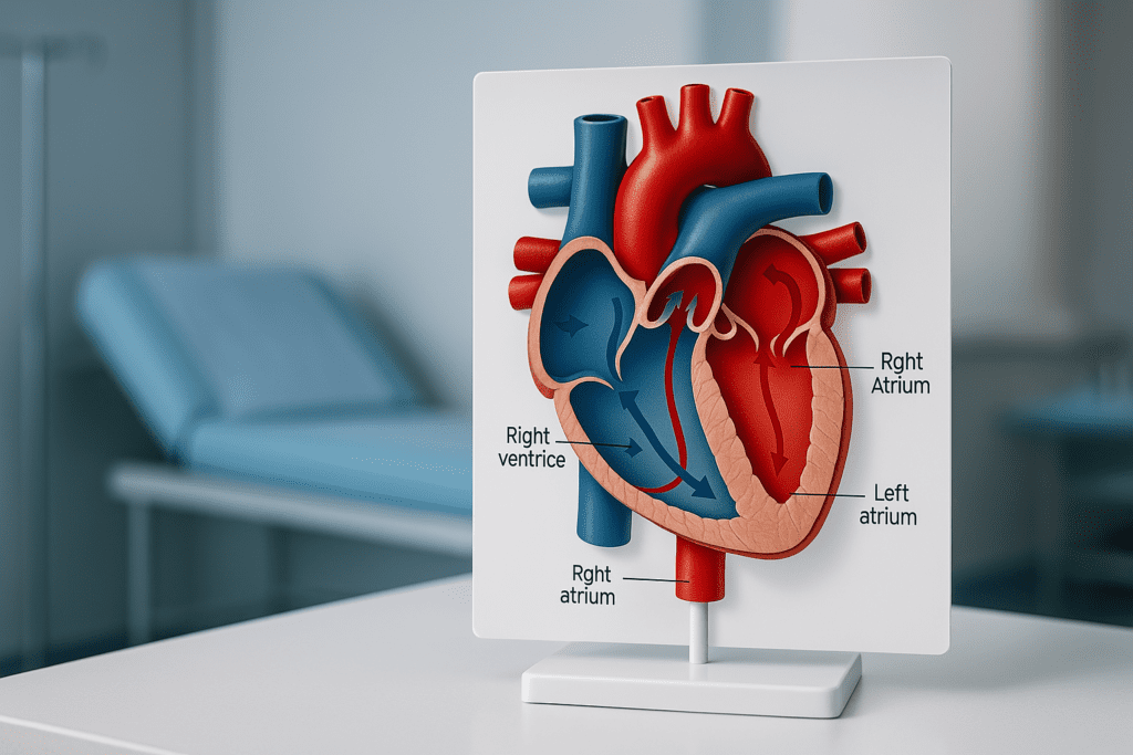
Pathway of Blood Through the Heart: A Step-by-Step Journey
To understand how the blood goes through the heart, it’s helpful to follow the path step by step, beginning with blood returning from the body. Deoxygenated blood enters the right atrium via the superior and inferior vena cava. From there, it moves through the tricuspid valve into the right ventricle. Upon ventricular contraction, the blood is pumped through the pulmonary valve into the pulmonary artery, which transports it to the lungs for oxygenation.
Once oxygenated in the lungs, blood returns to the left atrium via the pulmonary veins. It then flows through the mitral valve into the left ventricle. During systole, the left ventricle contracts and propels the blood through the aortic valve into the aorta, the body’s largest artery. From the aorta, oxygen-rich blood is distributed to all tissues via the arterial system. This complete cycle represents the order that blood flows through the heart and constitutes the route of blood through the heart from entry to exit.
Understanding this pathway helps answer questions like, “How does blood flow through the heart?” and “What is the order that the heart bumps blood flow?” These are not just academic queries but essential concepts in understanding human physiology. When reviewing a flow of heart diagram, each arrow or labeled chamber corresponds to a specific phase in this process, reinforcing the sequential nature of cardiac function.
Cardiac Anatomy: Structures Involved in Blood Flow
Each structure in the heart plays a critical role in ensuring unidirectional, efficient blood flow. The atria act as receiving chambers, while the ventricles function as high-pressure pumps. The four valves—tricuspid, pulmonary, mitral, and aortic—prevent backflow and maintain pressure gradients necessary for forward movement. The endocardium lines the chambers, while the myocardium provides contractile force. The pericardium, though not directly involved in blood movement, protects the heart and reduces friction during each beat.
Understanding which structure is part of the circulatory system helps clarify the roles each element plays. For instance, coronary arteries—often depicted in a detailed circulatory system diagram—supply the myocardium with oxygenated blood, a critical task considering the heart’s continuous workload. Meanwhile, major vessels such as the pulmonary artery and aorta serve as transitional points between the heart and the vascular system. When learning how the circulatory system works, it becomes apparent that these anatomical structures function in harmony to support systemic and pulmonary circulation.
The Role of Major Blood Vessels in Circulation
Vessels are essential components of the circulatory system. Arteries, veins, and capillaries each contribute to the efficient transport of blood. Arteries, such as the aorta and its branches, carry oxygenated blood away from the heart under high pressure. A common question arises: do arteries carry oxygenated blood? In systemic circulation, the answer is yes. However, in the pulmonary circuit, the pulmonary artery carries deoxygenated blood to the lungs, highlighting an important exception.
Veins, conversely, return blood to the heart. One might ask, “Which vessel travels toward the heart?” The answer is veins—specifically, the superior and inferior vena cava returning systemic blood and the pulmonary veins bringing oxygenated blood from the lungs. Capillaries form the bridge between arteries and veins, allowing exchange at the cellular level. Collectively, these vessels contribute to the blood flow chart through the body and form an indispensable part of the circulatory system.
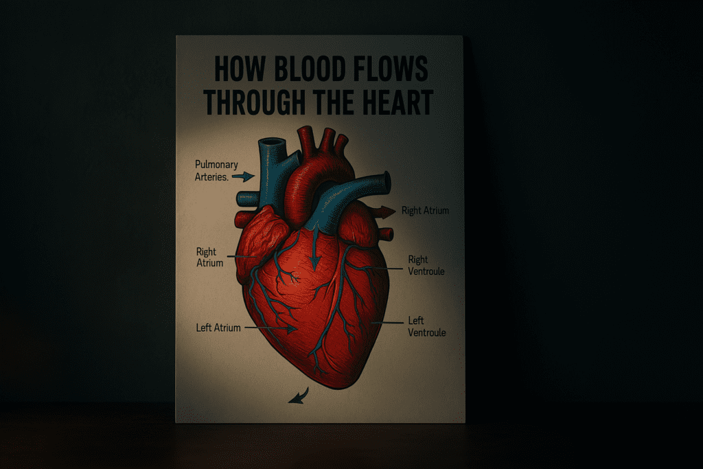
Blood Flow Diagrams and Their Educational Importance
Visual representations such as the blood flow in heart diagram or the cardiac blood flow diagram are essential tools for learning. These diagrams clearly map the sequential flow through each chamber and valve, reinforcing the textual description of the blood’s path. For those studying for heart system exams, these visuals serve as quick-reference guides, reducing cognitive load and increasing retention.
A typical blood flow chart through the body may incorporate both the pulmonary and systemic loops, showcasing the full circuit of blood from body to lungs and back. By using these diagrams, learners can identify each stage of the cycle, from atrial filling to ventricular ejection, and match these events with corresponding anatomical structures. Reviewing these images in tandem with textual explanations helps reinforce the natural order that blood flows through the heart and body, making them invaluable for both students and educators.
How the Circulatory System Works in Daily Life
The circulatory system is constantly adapting to the body’s needs. During exercise, for example, blood flow increases to skeletal muscles while temporarily decreasing to digestive organs. The heart responds to this demand by increasing its rate and contractility, a phenomenon known as cardiac output regulation. Understanding how the circulatory system works allows us to appreciate how seamlessly these adjustments are made, often without conscious awareness.
This functionality also has clinical relevance. Conditions such as heart failure, valvular disorders, or arrhythmias disrupt the normal pathway of blood through the heart, leading to symptoms like fatigue, shortness of breath, or edema. Recognizing these signs and understanding their basis in circulatory physiology empowers patients to seek care promptly. The question “What does the cardiovascular system do?” is not merely theoretical—it forms the basis for real-world clinical insight, diagnosis, and intervention.
Practical Insights for Students Studying the Heart System
For medical and allied health students, mastering the path of blood flow through the heart is a fundamental learning objective. Memorization strategies often involve tracing diagrams, using mnemonics, or rehearsing the sequence aloud. However, deeper learning arises from contextual understanding. Knowing not just the route but also the rationale—why valves open and close, how pressure changes guide flow—leads to a more meaningful grasp of cardiovascular physiology.
Resources such as animated models, 3D simulations, and guided dissection videos can enhance comprehension. These tools clarify not just how blood flows through the heart, but also how diseases alter this flow. Students preparing for exams must not only identify structures but also explain how they work together. The emphasis on functional integration is key when studying for heart system understanding, and it reinforces long-term retention over rote memorization.
Frequently Asked Questions: Advanced Insights Into Heart Function and Blood Flow
1. Why does blood flow direction matter in cardiac health?
The directionality of blood flow through the heart is essential for ensuring efficient oxygenation and nutrient distribution. If the order that blood flows through the heart is altered due to valve disease or structural defects, the body may suffer from inadequate perfusion, leading to fatigue, organ stress, or even heart failure. In clinical cardiology, one of the earliest signs of dysfunction is turbulent or regurgitant flow, which disrupts the standard path of blood flow through the heart and leads to decreased cardiac output. Accurate interpretation of a cardiac blood flow diagram helps physicians assess whether blood is moving properly through each chamber and valve. Without the correct route of blood through the heart, the circulatory system loses its ability to maintain stable internal conditions, which is critical for survival.
2. How does physical activity influence blood flow and heart performance?
Exercise causes dynamic adjustments in the cardiovascular system that improve the efficiency of blood flow through the heart. When we engage in sustained physical activity, the stroke volume increases, meaning more blood is pumped with each heartbeat. This adaptation enhances circulation without overburdening the heart. Over time, consistent aerobic activity strengthens the myocardium, reshaping the flow of blood through the heart by increasing ventricular efficiency. For those studying for heart system anatomy, understanding the way exercise impacts flow patterns and heart chamber dimensions is essential for recognizing the long-term benefits of cardiovascular fitness.
3. Are there structural variations in the circulatory system among individuals?
Yes, although the basic layout of the circulatory system is consistent, anatomical variations can exist. Some individuals are born with congenital anomalies such as a patent foramen ovale, bicuspid aortic valve, or even reversed organ placement (situs inversus), all of which may affect the pathway of blood through the heart. These variations require specific diagnostic approaches, often using a blood flow in heart diagram to detect deviations from normal. Knowing which structure is part of the circulatory system and how it functions in different configurations is vital for both diagnosis and treatment planning. Personalized medicine is increasingly adapting to these anatomical nuances to optimize cardiovascular care.
4. How do emotional and psychological states influence blood flow and heart function?
Emotions such as stress, anxiety, and excitement have a profound impact on blood flow and cardiovascular health. Stress triggers the sympathetic nervous system, releasing hormones like adrenaline that increase heart rate and contractility, thereby altering how the blood goes through the heart. Chronic emotional stress can lead to sustained hypertension and arrhythmias, affecting both the flow of blood through the heart and the broader circulatory system. Mental health thus becomes a critical factor in how the cardiovascular system performs over time. This interconnection underscores why heart health assessments should include discussions about emotional well-being, especially when interpreting a blood circulatory system diagram in clinical settings.
5. What advancements in imaging have improved our understanding of heart function?
Modern imaging technologies like 3D echocardiography, cardiac MRI, and CT angiography have revolutionized our ability to visualize the circulatory system. These tools provide detailed, dynamic blood flow chart through the body views, allowing clinicians to see real-time valve function and chamber pressures. Unlike traditional 2D images, newer modalities offer volumetric insights that clarify how blood flow through the heart changes with each heartbeat. For complex diagnoses, such as identifying blockages or assessing the route of blood through the heart, these imaging methods offer invaluable clarity. They also support patient education, turning abstract blood flow concepts into visible, understandable images.
6. What does the cardiac system do beyond moving blood?
Beyond its mechanical pumping role, the cardiac system integrates with hormonal and autonomic regulation networks. It participates in blood pressure modulation via baroreceptors and influences hormone distribution, such as antidiuretic hormone (ADH) and natriuretic peptides. This highlights that what the cardiac system does is more than just circulate blood—it actively communicates with other organs. Understanding how the heart works in this context means considering not only muscle contraction but also endocrine and neurological signals. The heart’s adaptability is showcased in every circulatory system diagram that maps its links to the brain, kidneys, and endocrine glands.
7. How do cardiovascular conditions disrupt the path of blood flow through the heart?
Diseases such as mitral regurgitation, aortic stenosis, or cardiomyopathy can severely alter the order that the heart bumps blood flow. For instance, in aortic stenosis, the narrowed valve obstructs the blood flow from the left ventricle to the aorta, reducing systemic circulation. A carefully interpreted cardiac blood flow diagram can reveal how these disruptions change the efficiency of the system. For students or clinicians, tracing the altered flow through a blood flow in heart diagram enables better understanding of symptom patterns. These insights aid in designing treatments that restore normal flow, often using prosthetic valves or cardiac remodeling strategies.
8. Why is venous return critical to maintaining proper blood flow?
Venous return refers to the process of blood traveling back toward the heart, particularly into the right atrium. This step completes the circulation cycle and maintains preload, the stretch of cardiac muscle before contraction. When venous return is compromised—due to dehydration, prolonged immobility, or venous insufficiency—it reduces cardiac output and disrupts the blood flow chart through the body. Knowing which vessel travels toward the heart, such as the superior and inferior vena cava, is essential for evaluating circulatory adequacy. This is often overlooked in surface-level studies of how the circulatory system works, yet it plays a pivotal role in sustaining life.
9. Can nutrition influence how the blood flows through the heart?
Absolutely. Nutritional choices directly affect vascular health, blood viscosity, and endothelial function. Diets high in saturated fat or sodium contribute to arterial stiffness, increasing resistance against which the heart must pump—thus altering how blood flow thru heart chambers operates. Conversely, omega-3 fatty acids, leafy greens, and antioxidants improve vessel elasticity and lower inflammation. A well-balanced diet can preserve the route of blood through the heart and body by supporting optimal pressure gradients. This preventive approach is reflected in global health guidelines, which increasingly emphasize dietary interventions to protect the integrity of the circulatory system.
10. What are the long-term effects of chronic circulatory system imbalances?
Persistent disruptions in blood flow can lead to organ damage, cognitive decline, and reduced quality of life. Conditions such as chronic hypertension or heart failure distort the path of blood flow through the heart, often overloading one chamber and weakening cardiac efficiency. Over time, this misalignment may show in a distorted flow of heart diagram, illustrating enlarged chambers or bypassed valves. For those interested in what the circulatory system does over decades of life, it’s important to understand the cumulative effects of these imbalances. Preventive cardiology now focuses on early identification of subtle abnormalities in blood flow to halt progression and preserve overall health.
Conclusion: Understanding Blood Flow in the Heart and Circulatory System as the Foundation of Cardiovascular Health
Gaining a deep understanding of how blood flows through the heart is foundational to mastering cardiovascular health. By appreciating the intricacies of the path of blood flow through the heart, and by studying the interplay of heart chambers, valves, and vessels through both textual explanations and visual aids like a circulatory system diagram, readers can grasp how vital this system is to every aspect of life. Recognizing the main function of the circulatory system—as a regulator of oxygen delivery, nutrient transport, and waste removal—makes it clear why even minor disruptions can lead to significant health consequences.
Throughout this article, we’ve examined how the heart works, what the cardiac system does, and how blood flow is regulated through a combination of anatomical precision and physiological control. We’ve also clarified frequently asked questions, including “Do arteries carry oxygenated blood?” and “What does the heart do?”, all while providing a strong framework for those studying for heart system exams or managing cardiovascular conditions.
Whether you’re a student, patient, or healthcare provider, understanding the route of blood through the heart and body helps you see the cardiovascular system not as a static structure, but as a dynamic, responsive network. With this knowledge, the once-complex blood flow chart through the body becomes a coherent narrative—a story of life delivered one heartbeat at a time.
heart muscle function, cardiac cycle explained, anatomy of the heart, cardiovascular physiology, pulmonary circulation process, systemic circulation explained, heart valve function, chamber contraction in heart, oxygen transport in blood, heart rate regulation, cardiac output mechanics, role of ventricles, atrial function in heart, myocardial performance, circulation and oxygenation, understanding cardiac rhythm, blood vessel anatomy, capillary exchange process, cardiovascular system overview, diagnostic heart imaging
Further Reading:
Heart Anatomy, Function, and Blood Circulation
Disclaimer
The information contained in this article is provided for general informational purposes only and is not intended to serve as medical, legal, or professional advice. While MedNewsPedia strives to present accurate, up-to-date, and reliable content, no warranty or guarantee, expressed or implied, is made regarding the completeness, accuracy, or adequacy of the information provided. Readers are strongly advised to seek the guidance of a qualified healthcare provider or other relevant professionals before acting on any information contained in this article. MedNewsPedia, its authors, editors, and contributors expressly disclaim any liability for any damages, losses, or consequences arising directly or indirectly from the use, interpretation, or reliance on any information presented herein. The views and opinions expressed in this article are those of the author(s) and do not necessarily reflect the official policies or positions of MedNewsPedia.