


Understanding the complexity and elegance of the human digestive system is essential for anyone who values nutrition, optimal wellness, and lifelong health. Far from being a simple biological process, digestion is a meticulously orchestrated function that involves a wide range of organs, hormones, enzymes, and biochemical pathways. When we examine the digestive tract labeled and fully illustrated in anatomical detail, we begin to appreciate how every meal sets off a dynamic cascade of events, each integral to our survival. From chewing food to absorbing nutrients at the microscopic level, the journey of digestion is as fascinating as it is vital. This article offers an in-depth exploration of how the human digestive system works, providing a scientifically sound yet reader-friendly perspective complete with labeled diagrams and expert insights for those interested in better nutrition and wellness.
You may also like: Macronutrients vs Micronutrients: What the Simple Definition of Macronutrients Reveals About Your Diet and Health
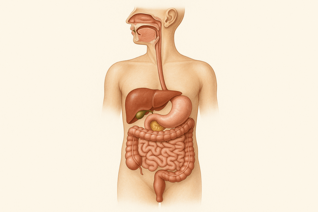
A Brief Overview of the Human Digestive System Diagram
The human digestive system is a continuous tube stretching from the mouth to the anus. When studying any human digestive system diagram, one immediately notices the interconnectedness of its parts. Each component plays a specialized role, and no part functions in isolation. A properly labeled diagram of the digestive system of man will usually include the mouth, pharynx, esophagus, stomach, small intestine, large intestine, rectum, and anus. Supporting organs such as the liver, pancreas, and gallbladder are also essential even though they are not directly part of the gastrointestinal tract. These accessory organs produce enzymes and bile that assist in breaking down macronutrients like fats, proteins, and carbohydrates. By examining a GI tract labeled visually and anatomically, it becomes easier to grasp the sequence of digestion and the symbiotic relationships among the system’s structures.
Mouth and Salivary Glands: The Unsuspected Power of Chewing
Digestion begins in the mouth, where mechanical and chemical processes initiate the breakdown of food. Chewing physically reduces food into smaller particles, while saliva—produced by the salivary glands—introduces enzymes like amylase that start digesting starches. This is more than just a preparatory step; it plays a pivotal role in signaling the rest of the digestive system to prepare for incoming nutrients. Many underestimate the significance of this early stage, but the anatomy simple digestive system cutaway images show the importance of salivary glands and their ducts feeding into the oral cavity. These glands help maintain oral health, lubricate food for swallowing, and trigger the release of digestive hormones. Looking at any simple digestive system diagram outline, we can observe how even the act of chewing has physiological ramifications throughout the digestive tract.
Esophagus and Peristalsis: The Silent Conveyor
Once food is chewed and mixed with saliva, it forms a soft mass called a bolus. Swallowing moves the bolus into the esophagus, a muscular tube that connects the throat to the stomach. The esophagus may seem like a passive structure at first glance, but a diagram of the human digestive tract reveals the unique longitudinal and circular muscle layers that facilitate peristalsis—the rhythmic contractions that push food downward. This movement is both involuntary and vital for moving food regardless of body position. Disorders that affect peristalsis can lead to significant discomfort and malnutrition. By analyzing a digestive process diagram, one can appreciate how the esophagus plays a crucial intermediary role in guiding food along its path.
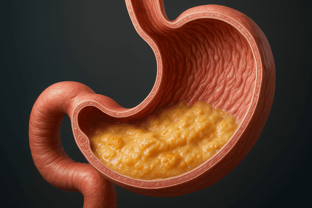
The Stomach: A Cauldron of Chemical Breakdown
The stomach is where the most aggressive chemical processing begins. Stomach acid (hydrochloric acid) and enzymes like pepsin break down proteins into smaller peptides. The anatomy of the stomach, shown in any comprehensive picture of digestive system processes, includes the fundus, body, antrum, and pylorus. Each region has a unique role, and their lining secretes various substances essential to digestion. For instance, intrinsic factor, crucial for vitamin B12 absorption, is produced in the stomach. A well-illustrated human digestive system labeled in detail will highlight not just the stomach’s location but also its inner lining and secretory functions. Furthermore, stomach contractions help churn food into a semi-liquid substance called chyme, which prepares it for the next stage of digestion.
Small Intestine: The Engine of Absorption
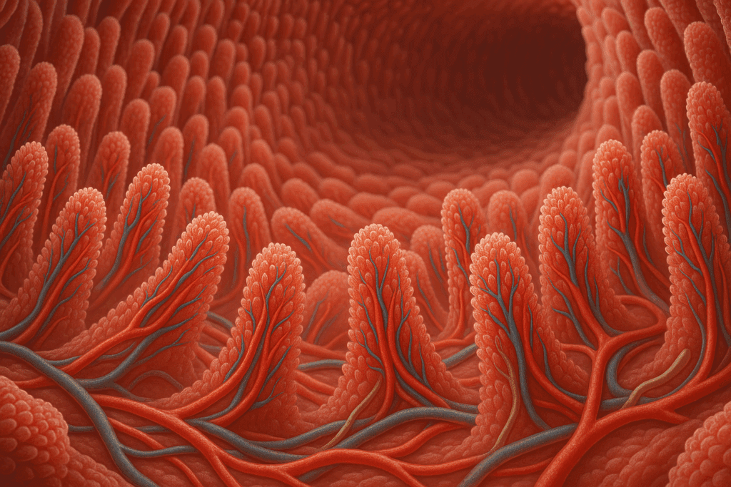
The small intestine is arguably the most critical part of the digestive tract for nutrient absorption. It consists of three segments: the duodenum, jejunum, and ileum. A detailed human digestive system diagram typically showcases how these segments differ in structure and function. The duodenum receives bile from the liver and enzymes from the pancreas, allowing for the emulsification of fats and further breakdown of proteins and carbohydrates. The jejunum and ileum are lined with villi and microvilli—finger-like projections that vastly increase surface area for absorption. Through these specialized structures, nutrients are absorbed into the bloodstream and lymphatic system. Understanding how this section functions can be greatly enhanced by reviewing a labeled diagram of the digestive system in humans that includes these microscopic anatomical features.
Large Intestine: Water Reclamation and Gut Health
After nutrients are absorbed in the small intestine, the remaining waste enters the large intestine. This organ, sometimes referred to in a digestive tract labeled chart as the colon, plays a pivotal role in reclaiming water and electrolytes from indigestible food matter. It also houses trillions of gut microbiota that influence digestion, immune function, and even mental health. A diagram of digestion often illustrates the large intestine as a segmented tube looping around the abdomen. The ascending, transverse, descending, and sigmoid sections all contribute to the reabsorption process and the formation of feces. Moreover, the appendix, once considered a vestigial organ, is now recognized for its immune functions. When reviewing a human digestive system diagram, the positioning of the colon and its bacterial colonies should not be overlooked, as they are central to wellness and disease prevention.
Rectum and Anus: The Final Act of Elimination
The rectum and anus are the concluding sections of the digestive system. They are responsible for the storage and controlled elimination of waste material. Although often omitted from simpler illustrations, any medically accurate diagram of the human digestive tract or digestive system labeling chart will include these structures. The rectum serves as a holding area until stretch receptors signal the need to defecate. The anus, with its internal and external sphincters, allows for voluntary control of bowel movements. This final stage, though often given little attention, is essential for maintaining internal homeostasis. Problems in this area, such as hemorrhoids or colorectal cancer, can significantly impair quality of life, reinforcing the need to understand every element depicted in a fully labeled digestive system diagram.
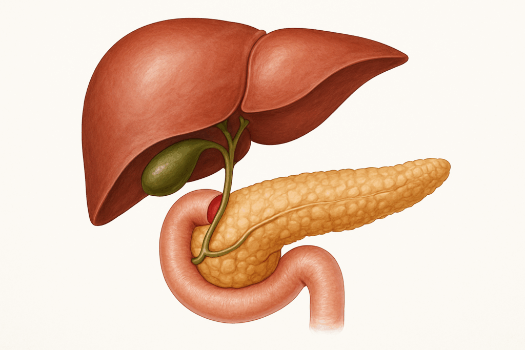
Accessory Organs: Silent Partners in Digestion
Though not part of the GI tract, the liver, gallbladder, and pancreas are integral to digestion. The liver produces bile, a substance crucial for emulsifying fats. The gallbladder stores and releases this bile in response to signals from the small intestine. The pancreas produces a cocktail of enzymes that break down carbohydrates, proteins, and fats, and it also secretes bicarbonate to neutralize stomach acid in the duodenum. These organs are consistently highlighted in any high-quality intestinal tract diagram due to their critical roles. Their interaction with the primary digestive organs underscores the systemic nature of digestion, where even distant organs contribute to nutrient processing. Looking at a comprehensive picture of digestive system anatomy makes their involvement abundantly clear.
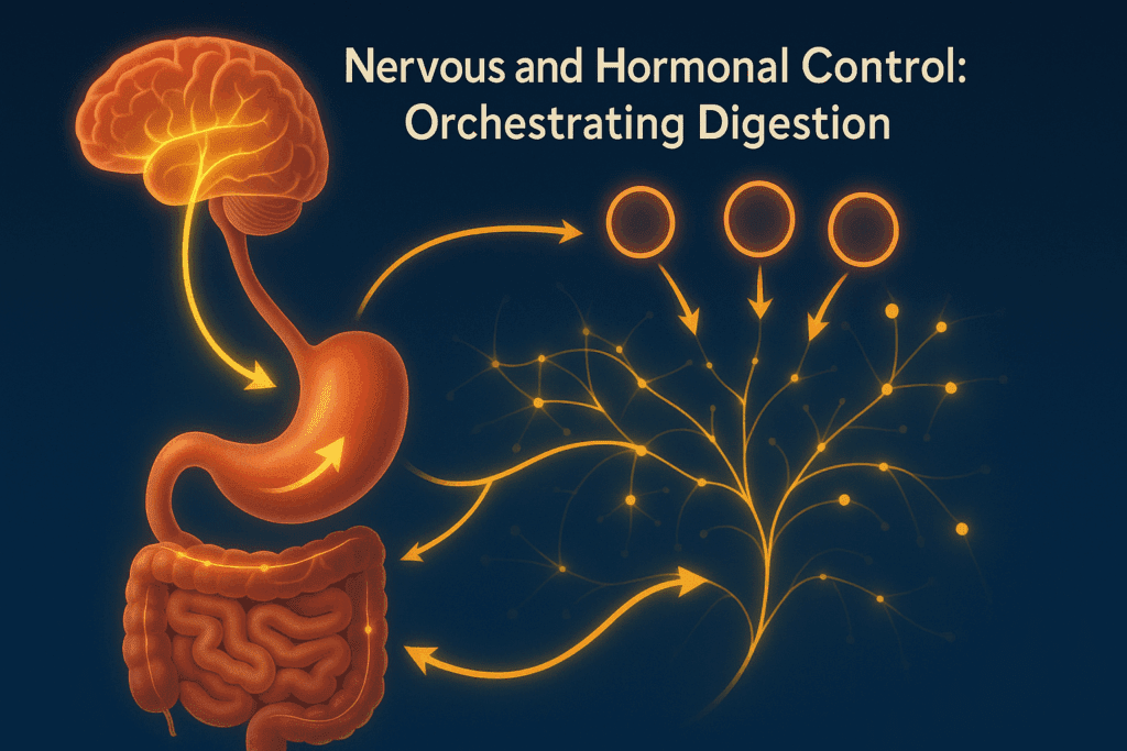
Nervous and Hormonal Control: Orchestrating Digestion
The digestive system operates under a finely tuned balance of neural and hormonal signals. The enteric nervous system, sometimes called the “second brain,” governs the local control of digestive activities, including peristalsis and enzyme secretion. Hormones such as gastrin, cholecystokinin (CCK), and secretin play pivotal roles in regulating digestive functions. When food enters the stomach, for example, gastrin stimulates acid production. As chyme moves into the duodenum, secretin and CCK are released, coordinating the release of bile and pancreatic enzymes. Any simple digestive system diagram outline is incomplete without recognizing this neurochemical overlay that choreographs digestion from start to finish. While not visible in a static diagram, their influence permeates every stage of the digestive process.
Digestive Health and Modern Nutrition
Understanding digestion provides critical insights into modern nutritional science. When we examine a detailed picture of the digestive system, it becomes clear why fiber is so important—it regulates bowel movement, feeds beneficial gut bacteria, and supports the health of the large intestine. Similarly, learning how fats are emulsified helps us understand the need for healthy fat sources and moderate intake. Visualizing the human digestive system labeled in an educational format can influence healthier dietary choices, from meal timing to portion control. It also brings clarity to common issues such as acid reflux, indigestion, or nutrient deficiencies. This knowledge supports both prevention and wellness, helping individuals align their diets with the natural design and needs of the digestive system.
Diagnostic Tools and Medical Imaging
The use of diagrams extends beyond education into diagnostic medicine. Tools like endoscopy, colonoscopy, and contrast imaging allow clinicians to visualize the digestive system in ways akin to a labeled diagram of digestion. These procedures often confirm findings suggested by anatomical diagrams, validating their accuracy and utility. Radiologists and gastroenterologists rely on visuals not unlike a human digestive system labeled illustration to interpret symptoms and prescribe treatment. Understanding these tools can empower patients to better grasp their diagnoses, comply with treatments, and engage in proactive health decisions.
Educational Impact of Digestive System Labeling
For educators and students alike, the ability to label the digestive system diagram is foundational to learning human biology. It’s not just about memorization, but about internalizing the function and purpose of each organ. This is why terms like digestive system in humans diagram and diagram of the digestive system of man remain popular in academic settings. Mastery of these labeled visuals cultivates a deeper understanding of bodily processes, which is particularly important for those entering health professions. A well-labeled and thoughtfully designed digestive system diagram serves as both a learning and teaching tool that transcends disciplines—from biology to clinical medicine to nutrition science.
Frequently Asked Questions: The Human Digestive System and Nutritional Wellness
1. How can understanding a labeled digestive system diagram help with dietary planning? Visualizing the human digestive system labeled in detail offers more than just academic value—it can directly influence how we approach food and nutrition. For example, knowing where the stomach ends and the small intestine begins helps in understanding the timing of nutrient absorption, which can affect how and when you eat. Athletes and individuals managing diabetes often use such insights to schedule meals that align with digestive efficiency. A well-labeled diagram of the digestive system of man can also highlight how specific nutrients are absorbed in different areas, guiding choices about supplements or nutrient-dense foods. When combined with an accurate digestion process diagram, this knowledge can transform a generic diet into a personalized wellness plan.
2. Are there psychological benefits to understanding digestion through labeled diagrams? Yes, studies suggest that individuals who understand their bodily processes often feel more empowered and less anxious about their health. A simple digestive system diagram outline or a full GI tract labeled image allows for better communication with healthcare providers, increasing confidence during consultations. For people with gastrointestinal issues such as IBS or GERD, having a clear picture of the digestive system can reduce the uncertainty that often exacerbates symptoms. Furthermore, educational tools like the anatomy simple digestive system cutaway can be especially calming for children and adults with health-related anxiety, helping them visualize normal function and reduce fear. In this way, a digestive tract labeled for clarity becomes a bridge between knowledge and emotional wellness.
3. Can digestive system labeling support early disease detection? Absolutely. When patients can accurately label the digestive system diagram, they often describe symptoms more clearly, aiding early diagnosis. For example, distinguishing between upper abdominal discomfort (often related to the stomach or duodenum) and lower abdominal pain (associated with the colon or rectum) becomes easier with reference to a human digestive system diagram. Early detection of conditions like Crohn’s disease, celiac disease, or even appendicitis can hinge on this understanding. Healthcare professionals increasingly recommend patient education involving the use of a labeled picture of digestive system structures during wellness visits. As medical literacy improves, so does the potential for prevention and timely intervention.
4. How is the intestinal tract diagram used in professional health settings? Medical professionals regularly refer to the intestinal tract diagram when discussing procedures like colonoscopies, endoscopies, or surgeries. For example, explaining the pathway of a colonoscope becomes easier with a digestive system in humans diagram, making the process more transparent for patients. Dietitians also use these diagrams to illustrate how fiber supports movement through the colon or how different foods affect the microbiome in specific areas of the large intestine. Additionally, these diagrams often appear in rehabilitation programs for patients recovering from gastrointestinal surgeries or bariatric procedures. The use of a diagram of the human digestive tract improves comprehension, adherence, and patient outcomes across disciplines.
5. What are some overlooked insights gained from a human digestive system labeled diagram? One lesser-known insight is the anatomical proximity between digestive and non-digestive organs. For instance, the pancreas not only plays a role in digestion but is also close to critical blood vessels and the spleen—knowledge that becomes vital during surgical planning. A properly labeled GI tract labeled chart can also reveal how closely bile ducts are situated to the liver and duodenum, explaining complications in gallstone diseases. Furthermore, a digestion process diagram can visually explain why certain medications need to be taken with food to avoid irritating the stomach lining. These nuanced insights underscore the value of comprehensive digestive system labeling in both personal and clinical contexts.
6. How do digital models of the digestive tract differ from textbook diagrams? While textbook versions like a traditional diagram of the digestive system of man offer foundational clarity, digital models provide dynamic, real-time visualization. Interactive tools allow users to manipulate a 3D digestive tract labeled model, observing how food moves through each section. These models often include animations that show muscle contractions, enzymatic reactions, and even microbiome activity. Compared to a static human digestive system diagram, digital resources enhance spatial awareness and interactivity, which can be particularly valuable for medical students or curious patients. These platforms sometimes integrate artificial intelligence to simulate diseases or nutrient deficiencies, offering a more immersive learning experience than a simple digestive system diagram outline.
7. Are there cultural or educational barriers to understanding a labeled digestive system diagram? Unfortunately, yes. In many parts of the world, limited access to quality science education makes it difficult for people to interpret a picture of digestive system structures accurately. Even in developed regions, language barriers can prevent patients from benefiting fully from diagrams unless they are available in multiple languages. Medical illustrations such as the anatomy simple digestive system cutaway often rely on prior knowledge of biological terminology, making them less accessible to individuals without a science background. For that reason, visual aids like the digestive system in humans diagram should be accompanied by clear, layperson-friendly explanations. Bridging this gap can empower more people globally to make informed dietary and medical decisions.
8. How does a better understanding of the GI tract labeled anatomy influence supplement usage? When individuals understand where and how nutrients are absorbed, they can make more informed choices about supplements. For example, iron is primarily absorbed in the duodenum, so time-released supplements that dissolve too late may be less effective. A diagram of digestion that highlights absorption sites allows users to align supplement timing and formulation with biological realities. People who refer to a digestive system labeling chart often discover why certain vitamins (like B12) require intrinsic factors or why probiotics must reach the colon intact to be effective. Understanding a digestive tract labeled by absorption phase transforms supplement use from guesswork into strategic action.
9. What are future innovations in digestive system visualization? Emerging technologies are making the traditional human digestive system diagram more interactive and personalized. For instance, augmented reality (AR) can now overlay a labeled diagram of the human digestive tract onto a person’s body, creating a real-time educational experience during medical consultations. Future applications might include wearable sensors that sync with apps to display a live GI tract labeled interface based on individual gut activity. As microbiome science evolves, diagrams may also highlight personalized bacterial colonies within the large intestine. The integration of machine learning into anatomy simple digestive system cutaway apps could simulate how lifestyle changes affect digestive function over time. These developments will transform passive viewing into dynamic learning.
10. How can educators make the topic of digestion more engaging using diagrams? Educators can enhance engagement by turning the act of labeling the digestive system diagram into an interactive, hands-on experience. Using color-coded cards, augmented reality, or even virtual dissection tools, students can build a physical or digital model of the digestive system in humans diagram. Field trips to anatomy labs or digital learning centers that display a 3D picture of digestive system pathways can make the content more tangible. Incorporating real-world applications, like tracking how a single meal moves through the digestive system, adds a narrative dimension that resonates with students. When students interact deeply with a diagram of digestion, they shift from passive memorization to active understanding, laying a foundation for lifelong nutritional awareness.
Conclusion: Why a Labeled Digestive System Diagram Matters for Wellness
The intricacies of digestion mirror the elegance of the human body’s inner workings. By taking the time to understand how each component of the digestive system functions—from the first bite of food to the final process of elimination—we empower ourselves to make smarter dietary and wellness decisions. Whether you’re a student learning to label the digestive system diagram or a health-conscious individual exploring the anatomy simple digestive system cutaway illustrations, the knowledge is profoundly impactful. When we observe a human digestive system labeled with precision or study a comprehensive intestinal tract diagram, we begin to align our nutrition with our biology. This alignment is the foundation of preventive health and nutritional wellness. The digestive tract labeled in anatomical context is more than just a visual aid; it’s a roadmap to better health, guiding us to treat our bodies with the respect, nourishment, and understanding they deserve.
Further Reading:
Anatomy of organs of the digestive system and their functions.
Overview of the Digestive System