


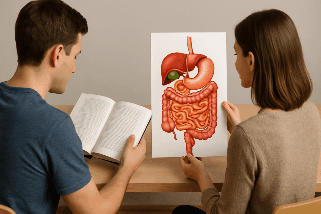
Understanding gut health requires more than simply knowing what foods to eat or which supplements to take. At its core, gut health is a dynamic interaction of complex biological systems, and one of the most effective ways to comprehend these systems is through visual learning. Digestive system images offer a profound window into the inner workings of the human gastrointestinal tract. From medical students to health-conscious adults, the use of a detailed digestive system photo can elevate the depth of understanding, bridging the gap between abstract anatomy and tangible health outcomes. In this guide, we explore how visual aids can enhance comprehension, promote preventive wellness, and support evidence-based learning in nutrition and digestive health.
You may also like: Macronutrients vs Micronutrients: What the Simple Definition of Macronutrients Reveals About Your Diet and Health
Why Visual Learning Matters in Digestive Health Education
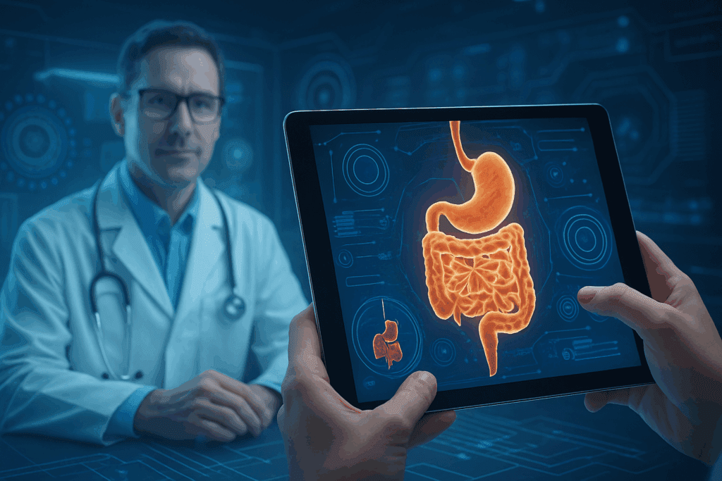
The human brain processes visual information exponentially faster than text. This neurological advantage becomes particularly important in health education, where dense medical terminology often obscures key concepts. When we pair visual aids such as digestive system images with scientific explanations, we enable a more intuitive grasp of anatomical structures, physiological processes, and potential disease states. Visuals don’t just supplement text—they reinforce it. For instance, a digestive system photo showing the esophagus, stomach, intestines, and accessory organs like the liver and pancreas provides an immediate sense of how food moves through the body. It contextualizes abstract descriptions and reinforces retention by linking visuals to real-world biological functions.
Medical educators have long recognized the value of anatomical images, but their relevance extends far beyond the classroom. For patients managing chronic gastrointestinal disorders, seeing a labeled digestive system photo during a consultation can make complex diagnoses easier to understand. This visual approach promotes informed decision-making, enhances doctor-patient communication, and reduces anxiety associated with health literacy challenges. By demystifying internal functions, visual tools empower individuals to actively engage in their own digestive health journey.
Unpacking the Digestive Process with Detailed Illustrations
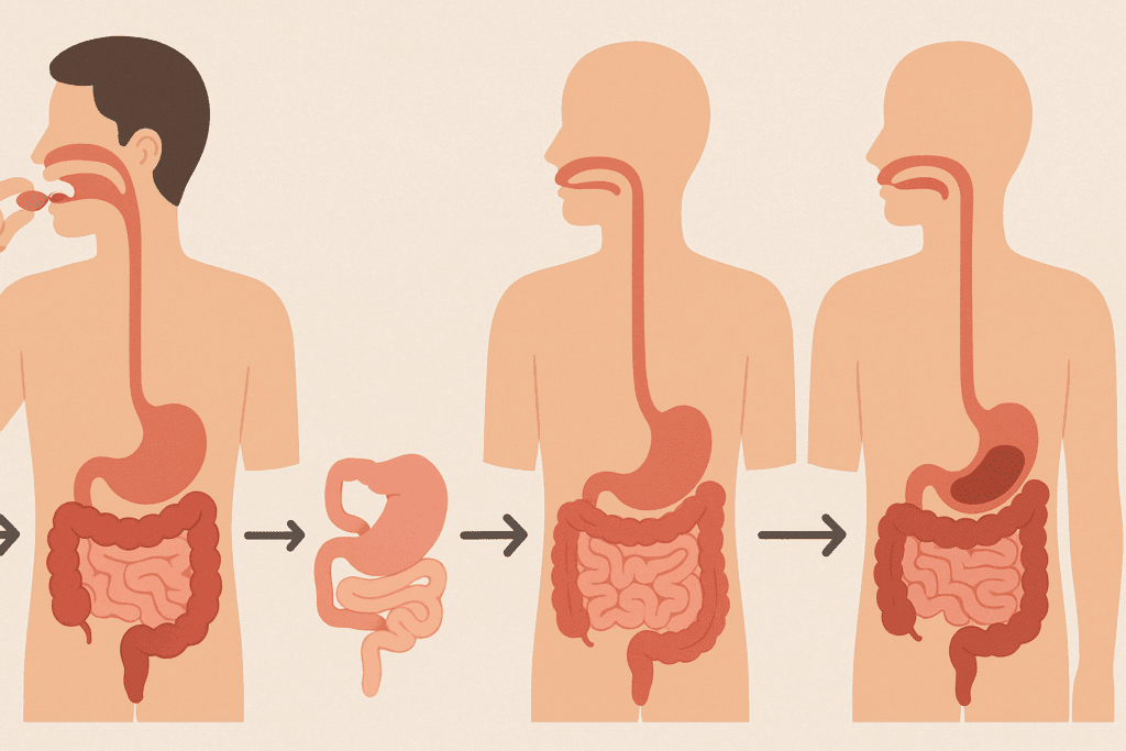
Digestive system images serve as more than just anatomical references; they map out the intricate sequence of digestion from ingestion to elimination. When paired with explanatory content, these visuals bring the mechanical and chemical stages of digestion to life. Take, for example, the process of mechanical breakdown in the mouth, followed by enzymatic action in the stomach and small intestine. A well-labeled digestive system photo can delineate each step clearly, showing how salivary glands, gastric acids, bile from the liver, and pancreatic enzymes contribute to nutrient breakdown.
Moreover, these illustrations help clarify how absorption occurs in the small intestine’s villi and how the large intestine reclaims water and electrolytes. The transition from nutrient extraction to waste formation can be abstract when described verbally, but a step-by-step visual progression makes it digestible—no pun intended. For individuals unfamiliar with terms like duodenum or jejunum, seeing these regions pinpointed in a digestive system image allows for immediate comprehension. It transforms theoretical anatomy into accessible, relatable content.
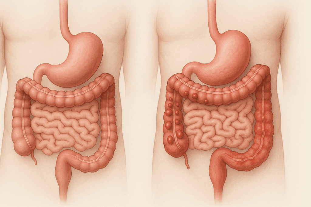
For educators and health communicators, the use of digestive system photos is essential when explaining pathophysiology. Illustrations that overlay normal function with abnormalities, such as ulcers, diverticula, or inflamed intestinal walls, offer comparative insight. These contrasts make it easier to grasp how disease disrupts normal digestion and why specific treatments or lifestyle changes are needed. When patients can visualize both wellness and dysfunction, they are more likely to adhere to prescribed regimens.
The Role of Digestive System Images in Nutritional Counseling
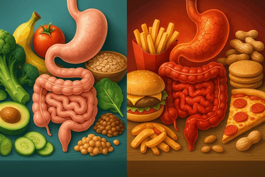
One of the most effective applications of digestive system images is in nutritional education. Dietary choices play a central role in digestive health, but their impact is often abstract. By integrating a digestive system photo into counseling sessions, nutritionists can illustrate the journey of specific food types and how they interact with gut physiology. For instance, a visual showing how dietary fiber bulks stool and accelerates intestinal transit provides more impact than a verbal explanation alone. It turns dietary advice into a visualized mechanism.
In cases of food intolerances or allergies, digestive system illustrations help patients understand which digestive organs are involved in processing certain foods and why symptoms such as bloating or diarrhea may occur. Seeing how the stomach produces enzymes to break down lactose or how gluten triggers inflammation in celiac disease within the small intestine reinforces the need for dietary restrictions. This visualization helps transform perceived food limitations into informed, health-empowering choices.
Furthermore, digestive system images can be powerful tools in promoting microbiome awareness. The gut microbiome—the diverse community of bacteria in the digestive tract—is increasingly recognized for its role in metabolism, immune function, and even mental health. Visual aids that depict microbial populations within the intestines help patients understand where probiotics act, how prebiotics nourish them, and why antibiotic overuse can be detrimental. These images clarify that gut health is not merely a function of digestion but a comprehensive system integral to total wellness.
Enhancing Disease Prevention with Digestive System Photo Education
Preventive health strategies depend on awareness and early intervention, and digestive system photos can serve as foundational educational tools in this context. When individuals understand the structure and function of their gastrointestinal systems, they are better equipped to recognize early warning signs of dysfunction. For example, understanding where the colon is located and how it functions can help individuals make sense of symptoms such as rectal bleeding or changes in bowel habits—both of which can signal conditions ranging from hemorrhoids to colorectal cancer.
In public health campaigns, visual resources that include detailed digestive system images have been shown to enhance engagement and improve message retention. These images provide a focal point for messaging about the importance of screening colonoscopies, liver function tests, or dietary interventions to prevent gallstones. When patients can see where these organs are situated and how they interact, the importance of preventive care becomes more tangible. Abstract concepts like “gastrointestinal health” become grounded in visible anatomy, fostering a sense of ownership and responsibility.
Additionally, digestive system images are instrumental in differentiating between normal aging-related changes and disease symptoms. An aging digestive system may experience slowed motility, decreased enzyme production, or changes in microbiome composition. By visually comparing youthful and aging digestive tracts, healthcare professionals can explain these physiological shifts more effectively. Patients gain reassurance about what to expect with age and are better able to distinguish when a symptom is part of normal aging versus a potential red flag.
Using Digestive System Images to Support Patient Empowerment
Visual education empowers patients, particularly when dealing with chronic gastrointestinal diseases such as irritable bowel syndrome (IBS), Crohn’s disease, or ulcerative colitis. These conditions can be confusing and emotionally taxing, especially when symptoms fluctuate or overlap. Digestive system images help patients visualize where inflammation or dysfunction is occurring and why certain medications target specific regions. For instance, a digestive system photo highlighting the ileum in Crohn’s disease can clarify why a treatment focuses on that area, fostering better understanding and adherence.
Patient empowerment also extends to self-care and lifestyle decisions. When individuals understand how digestion works, they are more likely to adopt behaviors that support gastrointestinal function, such as regular hydration, fiber intake, and mindful eating. Digestive system images reinforce the logic behind these practices, turning them from abstract health tips into science-backed habits. The act of visualizing these processes makes the connection between daily behavior and physiological response more direct and meaningful.
Moreover, for families and caregivers, especially of pediatric or elderly patients, these visuals can aid in explaining health conditions in ways that are accessible and respectful. Digestive system photos can be especially helpful when addressing sensitive topics like constipation, incontinence, or feeding tube placement, providing a non-threatening way to explore potentially embarrassing issues with clarity and compassion.
The Integration of Digestive System Photos in Digital Health Tools
As digital health platforms and mobile apps become increasingly prevalent, so too does the importance of integrating visual tools such as digestive system photos into user interfaces. From interactive anatomy modules to symptom-tracking tools, these visuals can improve user engagement and deepen understanding. For example, a mobile app that helps users monitor digestive symptoms can pair input fields with corresponding digestive system images to help users identify where discomfort is occurring. This improves accuracy in self-reporting and facilitates more productive discussions with healthcare providers.
Telemedicine platforms also benefit from the inclusion of digestive system images during virtual consultations. In the absence of in-person diagrams or physical models, a well-designed visual shared on-screen can clarify diagnoses and treatment plans, making telehealth interactions more effective. The ability to share and annotate digestive system photos in real time strengthens the therapeutic alliance and ensures patients leave virtual visits with a clear understanding of their health status.
Furthermore, augmented reality (AR) and virtual reality (VR) technologies are beginning to incorporate detailed digestive system illustrations to train medical students and educate patients. These immersive experiences provide a three-dimensional understanding of gastrointestinal anatomy and function, elevating the educational potential of traditional digestive system photos. As these technologies evolve, they hold the promise of making digestive health education more engaging, personalized, and accessible across diverse populations.

How Digestive System Images Foster Critical Thinking in Health Science
Beyond basic education, digestive system images foster analytical skills by encouraging users to connect visual data with theoretical knowledge. In academic settings, students learning physiology or pathology are often asked to interpret digestive system photos to identify abnormalities or trace the path of disease. This type of visual problem-solving sharpens diagnostic reasoning and reinforces understanding of normal versus abnormal processes.
For example, a case study involving abdominal pain may include a digestive system image for reference. Students are required to assess which regions correlate with the symptoms, drawing on both visual cues and textbook knowledge. This integrative learning style enhances long-term retention and mimics real-world clinical reasoning. The inclusion of such images in testing, training, and continuing education ensures that knowledge remains practical, not just theoretical.
This same approach applies to patient-facing materials. Brochures, educational websites, and hospital handouts that include digestive system photos allow patients to engage critically with their own health. Rather than passively receiving instructions, they can question, analyze, and understand the basis for treatment recommendations. This shared decision-making model—rooted in visual understanding—fosters collaboration and accountability.
Frequently Asked Questions (FAQ): How Digestive System Images Can Help You Understand Gut Health
1. How do digestive system images enhance the accuracy of symptom tracking for gastrointestinal conditions?
Digestive system images help individuals identify and localize symptoms more precisely, especially when describing discomfort during medical consultations. For instance, if someone experiences recurring pain in the lower right abdomen, a clear digestive system photo can assist them in visually pinpointing that region, which may correlate with the ileum or appendix. This specificity not only streamlines diagnosis but also empowers patients to better articulate their experiences. Many digital health apps now incorporate digestive system images to improve symptom logging accuracy, making them more useful for both patients and physicians. This enhanced communication can lead to quicker diagnosis, more appropriate testing, and a reduction in unnecessary procedures.
2. Can a digestive system photo help with understanding the role of lesser-known organs like the pancreas and gallbladder?
Absolutely. While the stomach and intestines are often the focus of gut health discussions, organs like the pancreas and gallbladder play crucial roles in digestion. A detailed digestive system photo offers an integrated view of how bile and digestive enzymes support fat and carbohydrate breakdown. Understanding the positioning and function of these organs through visual aids can help individuals recognize signs of dysfunction, such as gallbladder-related bloating or pancreatic enzyme insufficiency. Digestive system images also illustrate how these accessory organs connect with the rest of the gastrointestinal tract, making their roles in nutrient absorption more comprehensible.
3. Are there psychological benefits to using digestive system images in patient education?
Yes, using digestive system images in consultations can significantly reduce anxiety and confusion. Many patients feel overwhelmed when presented with complex medical explanations. However, when a doctor uses a digestive system photo to explain a diagnosis, it gives patients a visual anchor that enhances clarity and decreases fear. The psychological reassurance of being able to “see” the problem fosters trust and boosts adherence to treatment plans. Additionally, visual aids often increase perceived transparency and reduce the intimidation factor of medical jargon.
4. How are digestive system photos being adapted for pediatric health education?
Digestive system photos are increasingly being tailored for pediatric audiences, using child-friendly designs that simplify complex anatomical details without losing scientific integrity. These visuals are often colorful, interactive, and embedded in educational games or storybooks to teach children about gut health. By engaging young learners early, these images promote healthy habits like chewing thoroughly, eating fiber-rich foods, and drinking water. Pediatricians also report that digestive system images help children better understand what’s happening in their bodies when they experience symptoms like stomachaches or constipation. The ability to relate visual concepts to bodily sensations fosters emotional resilience and curiosity about health.
5. How do digestive system images support dietary planning for people with gastrointestinal disorders?
Individuals with conditions like IBS, GERD, or Crohn’s disease often need personalized dietary plans. Digestive system images can illustrate which parts of the gut are most affected by certain food triggers. For instance, those with GERD can benefit from a digestive system photo that shows the location and function of the lower esophageal sphincter, helping them understand why spicy or acidic foods may cause discomfort. Similarly, patients with Crohn’s can use visual aids to see why fiber intake needs to be adjusted during flare-ups. These images support the translation of clinical nutrition advice into understandable, actionable insights that patients can implement daily.
6. How are digestive system images being utilized in modern telehealth platforms?
As telehealth grows, integrating digestive system images into virtual consultations is becoming increasingly valuable. Doctors can share screen-annotated digestive system photos during video calls, allowing real-time visual explanations of diagnoses or treatment plans. This bridges the gap created by the lack of physical presence and compensates for the absence of in-office models or drawings. These tools enhance patient comprehension, especially for those with lower health literacy. Some advanced telehealth platforms are also incorporating interactive digestive system images, allowing patients to click on specific regions to learn about symptoms and treatment options, making digital healthcare more intuitive and informative.
7. Can using digestive system photos improve communication between caregivers and patients with dementia or cognitive impairment?
Yes, visual aids are especially useful for patients who struggle with verbal communication. A digestive system photo can act as a non-verbal tool that enables caregivers to ask clearer questions and interpret behaviors. For example, pointing to different regions of the digestive system image may help a patient with dementia indicate the source of their discomfort. This strategy enhances caregiving accuracy and reduces frustration on both sides. In long-term care settings, having accessible and labeled digestive system images can lead to earlier detection of digestive issues and prompt intervention, especially in populations at higher risk for complications due to age or cognitive decline.
8. What role do digestive system images play in raising public awareness about preventive screenings like colonoscopies?
Digestive system images are powerful visual aids in public health campaigns aimed at increasing colonoscopy rates and early detection of colorectal cancer. A clear and anatomically accurate digestive system photo can show the location of the colon, demystifying what the procedure examines and why it’s crucial. These visuals help address fears or misconceptions by breaking down the steps of the procedure in a digestible format. When patients can visualize where polyps form or where cancer typically starts, they are more likely to follow through with screenings. Public health organizations often incorporate digestive system images in brochures, posters, and social media to increase engagement and compliance.
9. How can digestive system photos aid in teaching gut-brain connection concepts?
While many people understand that digestion is physical, fewer grasp the neurological dimension of gut health. Digestive system photos that integrate elements of the nervous system can illustrate how the gut communicates with the brain via the vagus nerve and enteric nervous system. These enhanced visuals help explain why stress or anxiety can cause gastrointestinal symptoms and why gut inflammation can affect mood. The emerging science around the gut-brain axis becomes easier to comprehend when people can see the pathways involved. Using digestive system images in mental health or nutrition therapy sessions can bridge the knowledge gap between brain health and digestive function.
10. Are there future innovations in how digestive system images will be used in education or clinical care?
The future holds exciting developments in visual health education. Augmented reality (AR) and virtual reality (VR) are beginning to incorporate immersive digestive system images that allow users to “walk through” the GI tract in 3D. This can revolutionize medical training and enhance patient education, offering experiential learning instead of passive viewing. Artificial intelligence is also being integrated into these platforms to personalize digestive system image content based on individual health data. Eventually, digestive system photos may evolve into dynamic, interactive tools that adjust to a user’s symptoms, age, or disease history, making them not just informative but also diagnostic. These advancements will likely become standard in both clinical practice and public health education over the next decade.
Conclusion: Why Digestive System Images Matter for Gut Health and Lifelong Wellness
Digestive system images are not simply academic diagrams—they are essential tools that enhance understanding, improve communication, and support healthier lives. A well-chosen digestive system photo can demystify complex anatomy, clarify the effects of diet and disease, and serve as a bridge between scientific knowledge and practical wellness strategies. Whether viewed in a clinic, embedded in a mobile health app, or printed in an educational brochure, these visuals empower both professionals and patients to engage meaningfully with digestive health.
By integrating these images into educational, preventive, and clinical contexts, we promote a deeper understanding of the gastrointestinal system’s role in overall well-being. The ability to visualize where and how digestion occurs helps patients make informed choices, recognize potential warning signs, and adhere more confidently to treatment plans. Ultimately, digestive system photos are more than just illustrations—they are catalysts for health literacy, preventative action, and sustained wellness.
As our collective understanding of gut health continues to evolve, the role of visual education will only grow more important. For anyone committed to optimizing their digestive health, regularly consulting reliable, well-labeled digestive system images offers an invaluable starting point. With the guidance of healthcare professionals and the power of visual learning, we can foster a more informed, proactive, and healthy relationship with one of the body’s most vital systems.
Further Reading:
Your Digestive System in Pictures
Your Digestive System & How it Works