


Introduction: The Vital Role of the Human Heart and Its Position
The human heart is a vital organ responsible for sustaining life by pumping blood throughout the body. This complex muscular organ is crucial not only for circulation but for maintaining the body’s overall homeostasis. Its role in delivering oxygen and nutrients to tissues, while also removing waste products, makes understanding its location and anatomy essential for anyone studying the human body or dealing with cardiovascular health.
You may also like: 5 Ways to Keep Your Heart Healthy and Prevent Cardiovascular Disease
One of the first questions that often arises when discussing the heart is: Where is your heart located? While most people are aware that the heart is in the chest, its precise location and anatomical structure are much more complex than a simple answer reveals. Understanding the exact positioning of the heart within the body, along with its various chambers, vessels, and structures, provides crucial insights into how the heart functions. This knowledge is not only foundational for healthcare professionals but also important for individuals seeking to better understand their own cardiovascular health.
As we dive into the details of the heart’s location and anatomy, this article will explore the heart’s precise position in the chest, its various parts, and the intricate structures that make it function. By the end of this guide, readers will have a comprehensive understanding of the human heart’s anatomy, its position in the chest, and its role in maintaining life.
The Position of the Heart in the Body: Where Is It Located?
When most people are asked, “Where is your heart located?” they instinctively point to the left side of the chest. This is based on the common belief that the heart is predominantly situated on the left side of the body. While this is partly true, the full story is more nuanced.
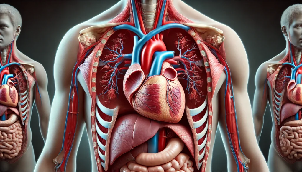
The human heart is located in the thoracic cavity, between the lungs in an area known as the mediastinum. It is roughly positioned behind the sternum, with its apex pointing to the left. However, this doesn’t mean that the entire heart lies exclusively on the left side of the body. In fact, the heart is slightly tilted, with approximately two-thirds of it residing on the left side of the body and the remaining third extending toward the right side.
The reason for this leftward tilt is due to the way the heart’s chambers and large vessels are arranged. The heart’s position can also vary slightly from person to person, influenced by factors like body size, posture, and even lung volume. For example, individuals with a larger chest cavity may have more space for the heart to shift, resulting in a slightly more central positioning.
Understanding the placement of the heart is not just a matter of anatomical curiosity—it also has important clinical implications. Cardiologists often examine the position of the heart when diagnosing conditions such as heart failure, arrhythmias, or pericarditis. Any abnormal shift in the heart’s position, such as displacement due to trauma or disease, can indicate significant medical concerns that may require immediate attention.
The Anatomy of the Human Heart: Key Components
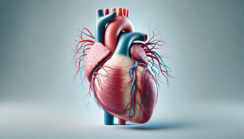
The human heart is not just a pump; it is a highly organized structure with distinct regions and parts that work together to perform its essential functions. Understanding heart anatomy involves exploring the heart’s chambers, valves, blood vessels, and surrounding structures. The human heart, in its entirety, is a muscular organ about the size of a fist. Its basic anatomy is composed of several key components that make it function optimally.
The Chambers of the Heart
The heart contains four distinct chambers: two atria (upper chambers) and two ventricles (lower chambers). The right atrium receives deoxygenated blood from the body via the superior and inferior vena cava, and pumps this blood into the right ventricle. The right ventricle then sends the blood to the lungs through the pulmonary artery for oxygenation. Once oxygenated, the blood returns to the left atrium via the pulmonary veins. The left atrium pumps the oxygen-rich blood into the left ventricle, which then distributes it throughout the body via the aorta.
This continuous cycle of blood flow is essential for the delivery of oxygen to tissues and the removal of carbon dioxide. Understanding how blood moves through these chambers—and the role each one plays—gives insight into how diseases such as heart failure, valve defects, or congenital heart conditions can disrupt normal circulation.
The Heart Valves: Gatekeepers of Blood Flow
The heart contains four essential valves that regulate blood flow, ensuring it moves in one direction. These valves are the tricuspid valve, pulmonary valve, mitral valve, and aortic valve. The tricuspid valve is located between the right atrium and the right ventricle, while the mitral valve is positioned between the left atrium and the left ventricle. The pulmonary valve controls blood flow from the right ventricle to the pulmonary artery, and the aortic valve regulates blood flow from the left ventricle to the aorta.
Each valve opens and closes in response to the pressures within the heart’s chambers. These valves are crucial for maintaining unidirectional blood flow and preventing backflow, which can lead to complications like regurgitation or valve stenosis. Understanding the structure and function of heart valves is integral to diagnosing and treating heart diseases such as valvular heart disease, which can cause serious disruptions to circulation.
Major Blood Vessels: Connecting the Heart to the Body
The heart is closely connected to the body through an intricate network of blood vessels that transport oxygenated and deoxygenated blood. These vessels include arteries, veins, and capillaries, all of which play a crucial role in circulation. The largest artery in the body, the aorta, emerges from the left ventricle and carries oxygen-rich blood to the entire body. The pulmonary arteries transport deoxygenated blood from the right ventricle to the lungs for oxygenation, while veins such as the superior and inferior vena cava carry deoxygenated blood from the body back to the right atrium.
These vessels form an interconnected system that ensures continuous blood flow to all areas of the body. Any blockage, narrowing, or dysfunction in these vessels can lead to severe complications such as a heart attack, stroke, or peripheral artery disease.
The Heart’s Location: Anatomical Insights into the Chest
The heart is positioned deep within the chest, specifically in the region between the two lungs. It rests slightly to the left of the sternum, with its apex pointing toward the left side of the body. In terms of vertical position, the heart is situated between the second and sixth ribs, with its base aligned near the center of the chest, just below the collarbones.
Understanding the heart’s position within the chest is not only important for anatomical studies but also for practical applications, such as medical imaging or surgical procedures. For instance, knowledge of heart placement is essential for performing accurate echocardiograms or CT scans, as well as for ensuring the safe insertion of medical devices like pacemakers or stents.
Frequently Asked Questions (FAQ) about Heart Anatomy
1. Where is the heart located in the human body?
The location of the heart in the human body is somewhat central, but it is tilted slightly to the left. It is situated behind the sternum (breastbone) and between the lungs in the chest cavity. Most people commonly associate the heart’s location with the left side of the chest, but it is actually slightly off-center. The heart is typically positioned with two-thirds of its mass on the left side of the body, and the remaining third on the right. If you’ve ever wondered, “Where is your heart located on a woman?” the anatomical heart anatomy doesn’t change based on gender, though it may appear slightly different in size or placement due to variations in body shape and size. Despite its tilted position, the heart’s function and placement remain crucial to its role in circulating blood throughout the body.
2. How many chambers does the human heart have?
The human heart consists of four chambers: two atria at the top and two ventricles at the bottom. These chambers work in sync to ensure the proper circulation of blood throughout the body. The right atrium receives deoxygenated blood from the body, while the left atrium receives oxygen-rich blood from the lungs. The right ventricle pumps the deoxygenated blood to the lungs for oxygenation, while the left ventricle pumps oxygenated blood throughout the rest of the body. This division of the heart into four chambers allows for efficient and controlled blood flow, maintaining the proper balance of oxygenated and deoxygenated blood in circulation. Understanding the chambers of the heart is essential when looking at heart health, as disruptions in this delicate balance can lead to various cardiovascular conditions.
3. What side is the heart on in the human body?
The heart is predominantly found on the left side of the chest, but it is more accurate to say that it is located near the center, with a slight tilt towards the left. While many people wonder, “What side is the heart on the body?” the heart’s position isn’t as simple as just being on the left or right. Instead, the heart’s placement in the chest cavity allows it to efficiently pump blood throughout the circulatory system. This positioning enables the left side of the heart, which handles oxygenated blood, to be closer to the lungs, where the oxygen is absorbed. Conversely, the right side of the heart is closer to the body’s extremities, making this arrangement essential for maintaining optimal blood circulation.
4. Where is your heart located on a woman’s body compared to a man’s?
The anatomical heart anatomy is largely the same in men and women, but there are some subtle differences. In terms of positioning, the heart’s location in a woman’s body is similar to a man’s, as it sits between the lungs in the chest cavity. However, women tend to have slightly smaller hearts than men due to differences in body size. This means that a woman’s heart may have a slightly higher heart rate than a man’s to compensate for the smaller size. Despite these differences, the heart placement in the body and its basic structure remain the same across genders, with the primary function of pumping oxygenated blood throughout the body. Whether in a heart model labeled for men or women, the essential anatomy and position of the heart are strikingly similar.
5. Is the heart located in the middle of the chest?
While the heart is near the center of the chest, it is not perfectly symmetrical or located exactly in the middle. Instead, the heart’s position in the chest is slightly leftward, with a majority of it located on the left side of the body. This slight tilt is why people typically feel the heartbeat more on the left side. If you were to look at a heart diagram or heart anatomy labeled with a ventral side view, you would see the left side of the heart is slightly larger and placed closer to the left lung. This asymmetry is crucial for the heart’s function in circulating blood through both the lungs and the rest of the body.
6. What parts of the heart are responsible for pumping blood to the body and lungs?
The heart consists of four main chambers that are responsible for circulating blood. The right side of the heart, specifically the right atrium and right ventricle, pumps deoxygenated blood to the lungs for oxygenation. The left side, made up of the left atrium and left ventricle, pumps oxygen-rich blood to the rest of the body. The left ventricle is the most powerful part of the heart as it has to push blood through the aorta and into the arteries, ensuring that oxygen reaches every part of the body. In terms of heart anatomy, the distinction between the right and left side is essential for understanding how blood flows through the heart and how the various chambers and valves function to regulate this flow.
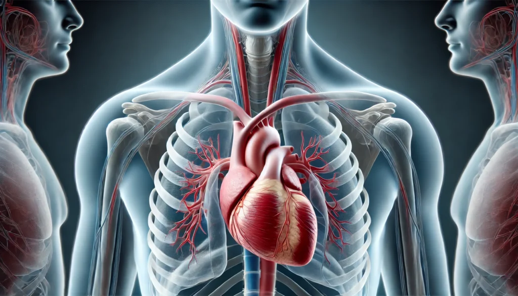
7. How does the heart’s position in the chest affect its function?
The heart’s placement in the chest plays a significant role in its ability to effectively pump blood throughout the body. Its central position, tilted slightly left, allows for the most efficient flow of blood to the lungs and to the rest of the body. The proximity of the heart to the lungs ensures that the right ventricle can pump blood to the lungs without excess distance. The location of the heart in the chest also helps protect it from physical trauma, as it is shielded by the rib cage. Additionally, this central positioning allows the heart to maintain a stable rhythm and efficient contraction, crucial for maintaining consistent circulation.
8. How big is the human heart compared to the rest of the body?
The human heart is relatively small compared to the body’s overall size. It weighs between 250 and 350 grams in most adults, which is less than 1% of your total body weight. Despite its small size, the heart is a powerhouse of activity, pumping roughly 70 milliliters of blood with every heartbeat. The size of the heart doesn’t change based on the side it’s on, whether left or right, but its ability to maintain blood flow is dependent on its capacity to contract forcefully and efficiently. Understanding the size and scale of the heart in relation to the rest of the body is important when considering conditions like heart failure, where the heart struggles to meet the body’s demand for oxygenated blood.
9. Can the heart’s location be different in some people?
In most people, the heart’s location remains consistent, but there are rare cases where the heart is positioned abnormally. In a condition known as situs inversus, the heart and other organs are mirrored, with the heart located on the right side of the chest instead of the left. This congenital condition doesn’t typically affect heart function, although it may require medical professionals to adjust their approach to diagnostics and treatment. However, for the vast majority of individuals, the heart’s location and position in the chest remain in a standard place, with the left side being the dominant side for blood circulation.
10. How can a heart diagram help me understand its anatomy better?
A heart diagram is an excellent tool for understanding the intricate details of heart anatomy. Labeled heart models, whether depicting the ventral side or showing a cross-section, allow you to visualize the heart’s structure. From the right atrium and ventricle to the left side with its atrium and powerful left ventricle, a labeled diagram helps clarify how blood flows through the heart. It can also provide insights into the heart’s valves and arteries, which play critical roles in blood circulation. Using a heart diagram labeled for anatomical study can be an indispensable tool in learning about the parts of the heart, its function, and the delicate balance required for effective circulatory health.
Conclusion: Appreciating the Complexity of the Heart’s Position and Anatomy
The human heart’s position and anatomy are central to its role in sustaining life. From its location in the chest to its intricate structure and chambers, every part of the heart plays a vital role in the body’s overall health and function. By understanding where the heart is located, its structure, and how it operates, individuals can gain a deeper appreciation for this remarkable organ and its impact on overall well-being.
Whether exploring the left or right side of the chest, considering how the heart’s chambers work together, or delving into its vital connection with blood vessels, the heart’s position and anatomy are far more complex than most people realize. This knowledge is invaluable not only for healthcare professionals but also for anyone looking to understand more about their own cardiovascular health.
heart health, cardiovascular anatomy, circulatory system, blood circulation, heart muscle, heart disease prevention, human circulatory system, cardiovascular wellness, oxygenated blood flow, heart rate regulation, heart function, heart protection, cardiovascular fitness, anatomy of the heart, left ventricle function, right ventricle circulation, heart valves, blood oxygenation, lung circulation, heart chamber anatomy, chest cavity anatomy
Further Reading:
Structure and Function of the Heart
Disclaimer
The information contained in this article is provided for general informational purposes only and is not intended to serve as medical, legal, or professional advice. While MedNewsPedia strives to present accurate, up-to-date, and reliable content, no warranty or guarantee, expressed or implied, is made regarding the completeness, accuracy, or adequacy of the information provided. Readers are strongly advised to seek the guidance of a qualified healthcare provider or other relevant professionals before acting on any information contained in this article. MedNewsPedia, its authors, editors, and contributors expressly disclaim any liability for any damages, losses, or consequences arising directly or indirectly from the use, interpretation, or reliance on any information presented herein. The views and opinions expressed in this article are those of the author(s) and do not necessarily reflect the official policies or positions of MedNewsPedia.